8BBM | pdb_00008bbm

DNA binding domain of J-DNA Binding Protein 1 (JBP1)


Experimental Data Snapshot

  • Method: X-RAY DIFFRACTION
  • Resolution: 1.95 Å
  • R-Value Free: 
    0.252 (Depositor), 0.261 (DCC) 
  • R-Value Work: 
    0.202 (Depositor), 0.206 (DCC) 
  • R-Value Observed: 
    0.205 (Depositor) 

Starting Model: experimental
View more details

wwPDB Validation 3D Report Full Report

Validation slider image for 8BBM

This is version 1.3 of the entry. See complete history

Literature

Distant sequence regions of JBP1 contribute to J-DNA binding.

de Vries, I.Ammerlaan, D.Heidebrecht, T.Celie, P.H.Geerke, D.P.Joosten, R.P.Perrakis, A.

(2023) Life Sci Alliance 6

  • DOI: https://doi.org/10.26508/lsa.202302150
  • Primary Citation Related Structures: 
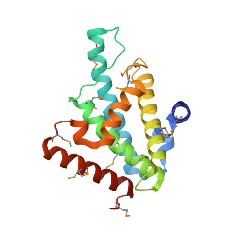
    8BBM

  • PubMed Abstract: 

    Base-J (β-D-glucopyranosyloxymethyluracil) is a modified DNA nucleotide that replaces 1% of thymine in kinetoplastid flagellates. The biosynthesis and maintenance of base-J depends on the base-J-binding protein 1 (JBP1) that has a thymidine hydroxylase domain and a J-DNA-binding domain (JDBD). How the thymidine hydroxylase domain synergizes with the JDBD to hydroxylate thymine in specific genomic sites, maintaining base-J during semi-conservative DNA replication, remains unclear. Here, we present a crystal structure of the JDBD including a previously disordered DNA-contacting loop and use it as starting point for molecular dynamics simulations and computational docking studies to propose recognition models for JDBD binding to J-DNA. These models guided mutagenesis experiments, providing additional data for docking, which reveals a binding mode for JDBD onto J-DNA. This model, together with the crystallographic structure of the TET2 JBP1-homologue in complex with DNA and the AlphaFold model of full-length JBP1, allowed us to hypothesize that the flexible JBP1 N-terminus contributes to DNA-binding, which we confirmed experimentally. Α high-resolution JBP1:J-DNA complex, which must involve conformational changes, would however need to be determined experimentally to further understand this unique underlying molecular mechanism that ensures replication of epigenetic information.


  • Organizational Affiliation
    • Oncode Institute and Division of Biochemistry, Netherlands Cancer Institute, Amsterdam, The Netherlands.

Macromolecule Content 

  • Total Structure Weight: 20.54 kDa 
  • Atom Count: 1,554 
  • Modeled Residue Count: 170 
  • Deposited Residue Count: 170 
  • Unique protein chains: 1

Macromolecules

Find similar proteins by:|  3D Structure
Entity ID: 1
MoleculeChains  Sequence LengthOrganismDetailsImage
Thymine dioxygenase JBP1170Leishmania tarentolaeMutation(s): 0 
Gene Names: JBP1
EC: 1.14.11.6
UniProt
Find proteins for Q9U6M1 (Leishmania tarentolae)
Explore Q9U6M1 
Go to UniProtKB:  Q9U6M1
Entity Groups
Sequence Clusters30% Identity50% Identity70% Identity90% Identity95% Identity100% Identity
UniProt GroupQ9U6M1
Sequence Annotations
Expand
Reference Sequence

Small Molecules

Modified Residues  1 Unique
IDChains TypeFormula2D DiagramParent
MSE
Query on MSE
A
L-PEPTIDE LINKINGC5 H11 N O2 SeMET

Experimental Data & Validation

Experimental Data

  • Method: X-RAY DIFFRACTION
  • Resolution: 1.95 Å
  • R-Value Free:  0.252 (Depositor), 0.261 (DCC) 
  • R-Value Work:  0.202 (Depositor), 0.206 (DCC) 
  • R-Value Observed: 0.205 (Depositor) 
Space Group: P 61 2 2
Unit Cell:
Length ( Å )Angle ( ˚ )
a = 68.286α = 90
b = 68.286β = 90
c = 185.842γ = 120
Software Package:
Software NamePurpose
REFMACrefinement
Aimlessdata scaling
PDB-REDOrefinement
XDSdata reduction
MOLREPphasing

Structure Validation

View Full Validation Report


Entry History 

& Funding Information

Deposition Data


Funding OrganizationLocationGrant Number
Netherlands Organisation for Scientific Research (NWO)Netherlands714.014.002

Revision History  (Full details and data files)

  • Version 1.0: 2022-11-09
    Type: Initial release
  • Version 1.1: 2023-07-05
    Changes: Database references
  • Version 1.2: 2024-02-07
    Changes: Data collection, Refinement description
  • Version 1.3: 2024-11-13
    Changes: Structure summary