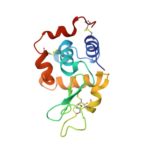
8AJ4

X-ray structure of lysozyme obtained upon reaction with [VIVO(malt)2] (Structure A')


Experimental Data Snapshot

  • Method: X-RAY DIFFRACTION
  • Resolution: 1.22 Å
  • R-Value Free: 0.203 
  • R-Value Work: 0.178 

Starting Model: experimental
View more details

wwPDB Validation   3D Report Full Report


Ligand Structure Quality Assessment 


This is version 1.2 of the entry. See complete history


Literature

Multiple and Variable Binding of Pharmacologically Active Bis(maltolato)oxidovanadium(IV) to Lysozyme.

Ferraro, G.Paolillo, M.Sciortino, G.Garribba, E.Merlino, A.

(2022) Inorg Chem 61: 16458-16467

  • DOI: https://doi.org/10.1021/acs.inorgchem.2c02690
  • Primary Citation of Related Structures:  
    8AJ3, 8AJ4, 8AJ5

  • PubMed Abstract: 

    The interaction with proteins of metal-based drugs plays a crucial role in their transport, mechanism, and activity. For an active ML n complex, where L is the organic carrier, various binding modes (covalent and non-covalent, single or multiple) may occur and several metal moieties (M, ML, ML 2 , etc.) may interact with proteins. In this study, we have evaluated the interaction of [V IV O(malt) 2 ] (bis(maltolato)oxidovanadium(IV) or BMOV, where malt = maltolato, i.e., the common name for 3-hydroxy-2-methyl-4 H -pyran-4-onato) with the model protein hen egg white lysozyme (HEWL) by electrospray ionization mass spectrometry, electron paramagnetic resonance, and X-ray crystallography. The multiple binding of different V-containing isomers and enantiomers to different sites of HEWL is observed. The data indicate both non-covalent binding of cis -[VO(malt) 2 (H 2 O)] and [VO(malt)(H 2 O) 3 ] + and covalent binding of [VO(H 2 O) 3-4 ] 2+ and cis -[VO(malt) 2 ] and other V-containing fragments to the side chains of Glu35, Asp48, Asn65, Asp87, and Asp119 and to the C-terminal carboxylate. Our results suggest that the multiple and variable interactions of potential V IV OL 2 drugs with proteins can help to better understand their solution chemistry and contribute to define the molecular basis of the mechanism of action of these intriguing molecules.


  • Organizational Affiliation

    Department of Chemical Sciences, University of Naples Federico II, Complesso Universitario di Monte Sant'Angelo, Via Cintia, I-80126Napoli, Italy.


Macromolecules
Find similar proteins by:  (by identity cutoff)  |  3D Structure
Entity ID: 1
MoleculeChains Sequence LengthOrganismDetailsImage
LysozymeA [auth AAA]129Gallus gallusMutation(s): 0 
EC: 3.2.1.17
UniProt
Find proteins for P00698 (Gallus gallus)
Explore P00698 
Go to UniProtKB:  P00698
Entity Groups  
Sequence Clusters30% Identity50% Identity70% Identity90% Identity95% Identity100% Identity
UniProt GroupP00698
Sequence Annotations
Expand
  • Reference Sequence
Small Molecules
Ligands 3 Unique
IDChains Name / Formula / InChI Key2D Diagram3D Interactions
MKO (Subject of Investigation/LOI)
Query on MKO

Download Ideal Coordinates CCD File 
D [auth AAA],
E [auth AAA],
F [auth AAA]
8,8-bis($l^{1}-oxidanyl)-2,2'-dimethyl-8,8'-spirobi[3$l^{4},7,9-trioxa-8$l^{6}-vanadabicyclo[4.3.0]nona-1(6),2,4-triene]
C12 H10 O8 V
KTNYRVZCCOTYPZ-UHFFFAOYSA-L
EPE
Query on EPE

Download Ideal Coordinates CCD File 
C [auth AAA]4-(2-HYDROXYETHYL)-1-PIPERAZINE ETHANESULFONIC ACID
C8 H18 N2 O4 S
JKMHFZQWWAIEOD-UHFFFAOYSA-N
NA
Query on NA

Download Ideal Coordinates CCD File 
B [auth AAA]SODIUM ION
Na
FKNQFGJONOIPTF-UHFFFAOYSA-N
Experimental Data & Validation

Experimental Data

  • Method: X-RAY DIFFRACTION
  • Resolution: 1.22 Å
  • R-Value Free: 0.203 
  • R-Value Work: 0.178 
  • Space Group: P 43 21 2
Unit Cell:
Length ( Å )Angle ( ˚ )
a = 77.08α = 90
b = 77.08β = 90
c = 38.09γ = 90
Software Package:
Software NamePurpose
REFMACrefinement
autoPROCdata reduction
autoPROCdata scaling
PHASERphasing

Structure Validation

View Full Validation Report


Ligand Structure Quality Assessment 


Entry History & Funding Information

Deposition Data


Funding OrganizationLocationGrant Number
Not funded--

Revision History  (Full details and data files)

  • Version 1.0: 2022-11-23
    Type: Initial release
  • Version 1.1: 2024-01-31
    Changes: Data collection, Refinement description
  • Version 1.2: 2024-10-23
    Changes: Structure summary