8ZAY | pdb_00008zay

Crystal structure of Fab from mouse monoclonal antibody 4A9 against Aquifex aeolicus RseP PDZ tandem


Experimental Data Snapshot

  • Method: X-RAY DIFFRACTION
  • Resolution: 2.30 Å
  • R-Value Free: 
    0.269 (Depositor), 0.269 (DCC) 
  • R-Value Work: 
    0.218 (Depositor), 0.217 (DCC) 
  • R-Value Observed: 
    0.220 (Depositor) 

Starting Model: experimental
View more details

wwPDB Validation 3D Report Full Report

Validation slider image for 8ZAY

This is version 1.1 of the entry. See complete history

Literature

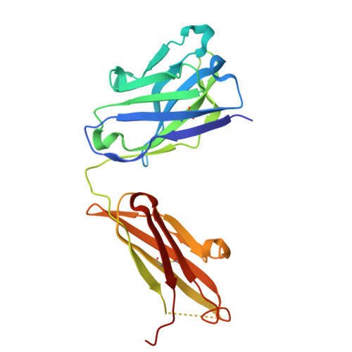
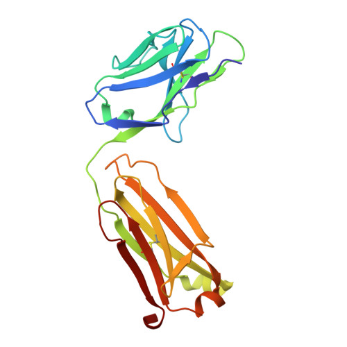
Cryo-EM structure of the bacterial intramembrane metalloprotease RseP in the substrate-bound state.

Asahi, K.Hirose, M.Aruga, R.Shimizu, Y.Tajiri, M.Tanaka, T.Adachi, Y.Tanaka, Y.Kaneko, M.K.Kato, Y.Akashi, S.Akiyama, Y.Hizukuri, Y.Kato, T.Nogi, T.

(2025) Sci Adv 11: eadu0925-eadu0925

  • DOI: https://doi.org/10.1126/sciadv.adu0925
  • Primary Citation Related Structures: 
    8ZAY, 9J82, 9J83

  • PubMed Abstract: 

    Site-2 proteases (S2Ps), conserved intramembrane metalloproteases that maintain cellular homeostasis, are associated with chronic infection and persistence leading to multidrug resistance in bacterial pathogens. A structural model of how S2Ps discriminate and accommodate substrates could help us develop selective antimicrobial agents. We previously proposed that the Escherichia coli S2P RseP unwinds helical substrate segments before cleavage, but the mechanism for accommodating a full-length membrane-spanning substrate remained unclear. Our present cryo-EM analysis of Aquifex aeolicus RseP ( Aa RseP) revealed that a substrate-like membrane protein fragment from the expression host occupied the active site while spanning a transmembrane cavity that is inaccessible via lateral diffusion. Furthermore, in vivo photocrosslinking supported that this substrate accommodation mode is recapitulated on the cell membrane. Our results suggest that the substrate accommodation by threading through a conserved membrane-associated region stabilizes the substrate-complex and contributes to substrate discrimination on the membrane.


  • Organizational Affiliation
    • Graduate School of Medical Life Science, Yokohama City University, 1-7-29 Suehiro-cho, Tsurumi-ku, Yokohama 230-0045, Japan.

Macromolecule Content 

  • Total Structure Weight: 96.85 kDa 
  • Atom Count: 6,893 
  • Modeled Residue Count: 863 
  • Deposited Residue Count: 880 
  • Unique protein chains: 2

Macromolecules

Find similar proteins by:|  3D Structure
Entity ID: 1
MoleculeChains  Sequence LengthOrganismDetailsImage
VH-CH1 region of antibody of anti-RseP orthologue from A aeolicusA [auth H],
B [auth I]
226Mus musculusMutation(s): 0 
Find similar proteins by:|  3D Structure
Entity ID: 2
MoleculeChains  Sequence LengthOrganismDetailsImage
Light chain of antibody of anti-RseP orthologue from A aeolicusC [auth L],
D [auth M]
214Mus musculusMutation(s): 0 

Small Molecules

Ligands 1 Unique
IDChains Name / Formula / InChI Key2D Diagram3D Interactions
ZN

Query on ZN


Download:Ideal Coordinates CCD File
E [auth H]
F [auth H]
G [auth I]
H [auth I]
I [auth L]
E [auth H],
F [auth H],
G [auth I],
H [auth I],
I [auth L],
J [auth L],
K [auth L],
L [auth M],
M,
N [auth M]
ZINC ION
Zn
PTFCDOFLOPIGGS-UHFFFAOYSA-N

Experimental Data & Validation

Experimental Data

  • Method: X-RAY DIFFRACTION
  • Resolution: 2.30 Å
  • R-Value Free:  0.269 (Depositor), 0.269 (DCC) 
  • R-Value Work:  0.218 (Depositor), 0.217 (DCC) 
  • R-Value Observed: 0.220 (Depositor) 
Space Group: P 1 21 1
Unit Cell:
Length ( Å )Angle ( ˚ )
a = 51.78α = 90
b = 70.44β = 96.69
c = 132.46γ = 90
Software Package:
Software NamePurpose
PHENIXrefinement
XDSdata reduction
XSCALEdata scaling
MOLREPphasing

Structure Validation

View Full Validation Report


Entry History 

& Funding Information

Deposition Data


Funding OrganizationLocationGrant Number
Japan Society for the Promotion of Science (JSPS)Japan23K23825
Japan Society for the Promotion of Science (JSPS)Japan22H02561

Revision History  (Full details and data files)

  • Version 1.0: 2025-03-05
    Type: Initial release
  • Version 1.1: 2025-03-12
    Changes: Database references, Structure summary