8Z55 | pdb_00008z55

Crystal structure of human SIRT5 in complex with succPrx1 and ADP ribose


Experimental Data Snapshot

  • Method: X-RAY DIFFRACTION
  • Resolution: 1.83 Å
  • R-Value Free: 
    0.246 (Depositor), 0.246 (DCC) 
  • R-Value Work: 
    0.212 (Depositor), 0.213 (DCC) 
  • R-Value Observed: 
    0.213 (Depositor) 

Starting Model: experimental
View more details

wwPDB Validation 3D Report Full Report

Validation slider image for 8Z55

Ligand Structure Quality Assessment 


This is version 1.0 of the entry. See complete history

Literature

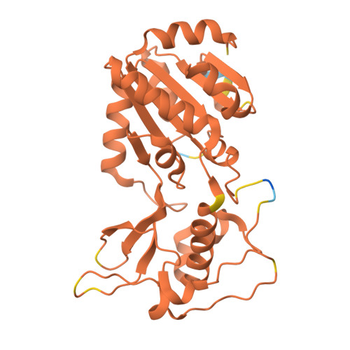
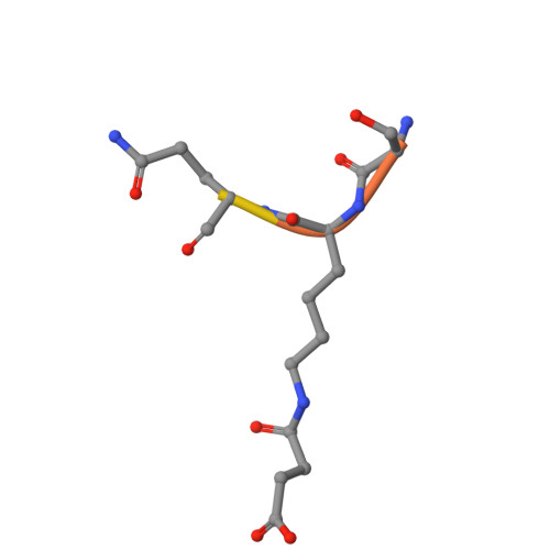
SIRT5 mutants reveal the role of conserved asparagine and glutamine residues in the NAD + -binding pocket.

Yokoyama, T.Takayama, Y.Mizuguchi, M.Nabeshima, Y.Kusaka, K.

(2024) FEBS Lett 598: 2269-2280

  • DOI: https://doi.org/10.1002/1873-3468.14961
  • Primary Citation Related Structures: 
    8Z54, 8Z55, 8Z56, 8Z57, 8Z58

  • PubMed Abstract: 

    SIRT5, one of the mammalian sirtuins, specifically recognizes succinyl-lysine residues on proteins and catalyzes the desuccinylation reaction. In this study, we characterized SIRT5 mutants with hydrophobic amino acid substitutions at Q140 and N141, in addition to the catalytic residue H158, known as an active site residue, by the Michaelis-Menten analysis and X-ray crystallography. Kinetic analysis showed that the catalytic efficiency (k cat /K m ) of the Q140L and N141V mutants decreased to 0.02 times and 0.0038 times that of the wild-type SIRT5, respectively, with the activity of the N141V mutant becoming comparable to that of the H158M mutant. Our findings indicate that N141 contributes significantly to the desuccinylation reaction.


  • Organizational Affiliation
    • Faculty of Pharmaceutical Sciences, University of Toyama, Japan.

Macromolecule Content 

  • Total Structure Weight: 31.75 kDa 
  • Atom Count: 2,232 
  • Modeled Residue Count: 270 
  • Deposited Residue Count: 283 
  • Unique protein chains: 2

Macromolecules

Find similar proteins by:|  3D Structure
Entity ID: 1
MoleculeChains  Sequence LengthOrganismDetailsImage
NAD-dependent protein deacylase sirtuin-5, mitochondrial274Homo sapiensMutation(s): 0 
Gene Names: SIRT5SIR2L5
EC: 2.3.1
UniProt & NIH Common Fund Data Resources
Find proteins for Q9NXA8 (Homo sapiens)
Explore Q9NXA8 
Go to UniProtKB:  Q9NXA8
PHAROS:  Q9NXA8
GTEx:  ENSG00000124523 
Entity Groups
Sequence Clusters30% Identity50% Identity70% Identity90% Identity95% Identity100% Identity
UniProt GroupQ9NXA8
Sequence Annotations
Expand
Reference Sequence
Find similar proteins by:  Sequence   |   3D Structure  
Entity ID: 2
MoleculeChains  Sequence LengthOrganismDetailsImage
Peroxiredoxin-1 fragment9Homo sapiensMutation(s): 0 
EC: 1.11.1.24
UniProt & NIH Common Fund Data Resources
Find proteins for Q06830 (Homo sapiens)
Explore Q06830 
Go to UniProtKB:  Q06830
PHAROS:  Q06830
GTEx:  ENSG00000117450 
Entity Groups
UniProt GroupQ06830
Sequence Annotations
Expand
Reference Sequence

Small Molecules

Modified Residues  1 Unique
IDChains TypeFormula2D DiagramParent
SLL
Query on SLL
B
L-PEPTIDE LINKINGC10 H18 N2 O5LYS

Experimental Data & Validation

Experimental Data

  • Method: X-RAY DIFFRACTION
  • Resolution: 1.83 Å
  • R-Value Free:  0.246 (Depositor), 0.246 (DCC) 
  • R-Value Work:  0.212 (Depositor), 0.213 (DCC) 
  • R-Value Observed: 0.213 (Depositor) 
Space Group: C 2 2 21
Unit Cell:
Length ( Å )Angle ( ˚ )
a = 46.198α = 90
b = 73.345β = 90
c = 163.938γ = 90
Software Package:
Software NamePurpose
PHENIXrefinement
XDSdata reduction
XSCALEdata scaling
MOLREPphasing

Structure Validation

View Full Validation Report


Ligand Structure Quality Assessment 


Entry History 

& Funding Information

Deposition Data


Funding OrganizationLocationGrant Number
Not funded--

Revision History  (Full details and data files)

  • Version 1.0: 2024-10-09
    Type: Initial release