8YXB

Crystal structure of the HSA complex with ceftriaxone and myristate


Experimental Data Snapshot

  • Method: X-RAY DIFFRACTION
  • Resolution: 2.20 Å
  • R-Value Free: 0.264 
  • R-Value Work: 0.233 
  • R-Value Observed: 0.235 

Starting Model: experimental
View more details

wwPDB Validation   3D Report Full Report


Ligand Structure Quality Assessment 


This is version 1.0 of the entry. See complete history


Literature

Interaction of Cephalosporins with Human Serum Albumin: A Structural Study.

Kawai, A.Yamasaki, K.Otagiri, M.Doi, Y.

(2024) J Med Chem 

  • DOI: https://doi.org/10.1021/acs.jmedchem.4c00983
  • Primary Citation of Related Structures:  
    8YXA, 8YXB

  • PubMed Abstract: 

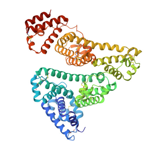
    Modification of the R1 and R2 side chain structures has been used as the main strategy to expand the spectrum of cephalosporins and impart resistance to hydrolysis by β-lactamases. These structural modifications also result in a wide range of plasma protein binding, especially with human serum albumin (HSA). Here, we determined the crystal structures of the HSA complexes with two clinically important cephalosporins, ceftriaxone and cefazolin, and evaluated the binding of cephalosporin to HSA by susceptibility testing and competitive binding assay. Ceftriaxone and cefazolin bind to subdomain IB of HSA, and their cephem core structures are recognized by Arg117 of HSA. Tyr161 of HSA changes its rotamer depending on the cephalosporin, resulting in alterations of the cavity shape occupied by the R2 side chain of cephalosporins. These findings provide structural insight into the mechanisms underlying the HSA binding of cephalosporins.


  • Organizational Affiliation

    Department of Microbiology, Fujita Health University School of Medicine, 1-98, Dengakugakubo, Kutsukake-cho, Toyoake, Aichi 470-1192, Japan.


Macromolecules
Find similar proteins by:  (by identity cutoff)  |  3D Structure
Entity ID: 1
MoleculeChains Sequence LengthOrganismDetailsImage
Serum albumin
A, B
585Homo sapiensMutation(s): 0 
Gene Names: ALBGIG20GIG42PRO0903PRO1708PRO2044PRO2619PRO2675UNQ696/PRO1341
UniProt & NIH Common Fund Data Resources
Find proteins for P02768 (Homo sapiens)
Explore P02768 
Go to UniProtKB:  P02768
PHAROS:  P02768
GTEx:  ENSG00000163631 
Entity Groups  
Sequence Clusters30% Identity50% Identity70% Identity90% Identity95% Identity100% Identity
UniProt GroupP02768
Sequence Annotations
Expand
  • Reference Sequence
Small Molecules
Ligands 2 Unique
IDChains Name / Formula / InChI Key2D Diagram3D Interactions
9F2 (Subject of Investigation/LOI)
Query on 9F2

Download Ideal Coordinates CCD File 
C [auth A],
I [auth B]
Ceftriaxone
C18 H18 N8 O7 S3
VAAUVRVFOQPIGI-TYHRLYECSA-N
MYR
Query on MYR

Download Ideal Coordinates CCD File 
D [auth A]
E [auth A]
F [auth A]
G [auth A]
H [auth A]
D [auth A],
E [auth A],
F [auth A],
G [auth A],
H [auth A],
J [auth B],
K [auth B],
L [auth B],
M [auth B],
N [auth B],
O [auth B]
MYRISTIC ACID
C14 H28 O2
TUNFSRHWOTWDNC-UHFFFAOYSA-N
Experimental Data & Validation

Experimental Data

  • Method: X-RAY DIFFRACTION
  • Resolution: 2.20 Å
  • R-Value Free: 0.264 
  • R-Value Work: 0.233 
  • R-Value Observed: 0.235 
  • Space Group: P 1
Unit Cell:
Length ( Å )Angle ( ˚ )
a = 38.893α = 105.73
b = 93.786β = 100.03
c = 94.747γ = 89.71
Software Package:
Software NamePurpose
PHENIXrefinement
XDSdata reduction
XDSdata scaling
MOLREPphasing

Structure Validation

View Full Validation Report


Ligand Structure Quality Assessment 


Entry History & Funding Information

Deposition Data

  • Released Date: 2024-08-07 
  • Deposition Author(s): Kawai, A.

Funding OrganizationLocationGrant Number
Japan Society for the Promotion of Science (JSPS)JapanJP20K16060

Revision History  (Full details and data files)

  • Version 1.0: 2024-08-07
    Type: Initial release