8YWP | pdb_00008ywp

Crystal structure of the Fab fragment of anti-IL-6 antibody I9H


Experimental Data Snapshot

  • Method: X-RAY DIFFRACTION
  • Resolution: 2.50 Å
  • R-Value Free: 
    0.285 (Depositor), 0.290 (DCC) 
  • R-Value Work: 
    0.258 (Depositor), 0.264 (DCC) 
  • R-Value Observed: 
    0.260 (Depositor) 

Starting Model: experimental
View more details

wwPDB Validation 3D Report Full Report

Validation slider image for 8YWP

This is version 1.2 of the entry. See complete history

Literature

Structural basis of signaling complex inhibition by IL-6 domain-swapped dimers.

Yudenko, A.Bukhdruker, S.Shishkin, P.Rodin, S.Burtseva, A.Petrov, A.Pigareva, N.Sokolov, A.Zinovev, E.Eliseev, I.Remeeva, A.Marin, E.Mishin, A.Gordeliy, V.Gushchin, I.Ischenko, A.Borshchevskiy, V.

(2025) Structure 33: 171-180.e5

  • DOI: https://doi.org/10.1016/j.str.2024.10.028
  • Primary Citation Related Structures: 
    8YWP, 8YWQ, 8YWR

  • PubMed Abstract: 

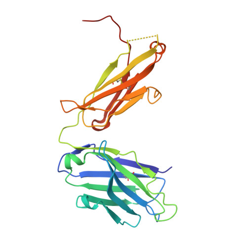
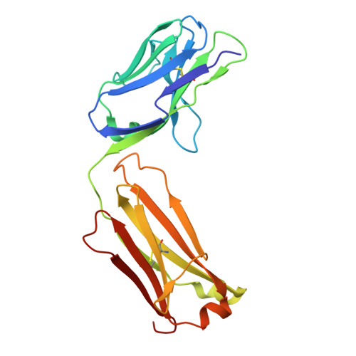
    Interleukin-6 (IL-6) is a multifaceted cytokine essential in many immune system processes and their regulation. It also plays a key role in hematopoiesis, and in triggering the acute phase reaction. IL-6 overproduction is critical in chronic inflammation associated with autoimmune diseases like rheumatoid arthritis and contributes to cytokine storms in COVID-19 patients. Over 20 years ago, researchers proposed that IL-6, which is typically monomeric, can also form dimers via a domain-swap mechanism, with indirect evidence supporting their existence. The physiological significance of IL-6 dimers was shown in B-cell chronic lymphocytic leukemia. However, no structures have been reported so far. Here, we present the crystal structure of an IL-6 domain-swapped dimer that computational approaches could not predict. The structure explains why the IL-6 dimer is antagonistic to the IL-6 monomer in signaling complex formation and provides insights for IL-6 targeted therapies.


  • Organizational Affiliation
    • Research Center for Molecular Mechanisms of Aging and Age-Related Diseases, Moscow Institute of Physics and Technology, Dolgoprudny, Moscow Region 141701, Russia.

Macromolecule Content 

  • Total Structure Weight: 48.64 kDa 
  • Atom Count: 3,505 
  • Modeled Residue Count: 440 
  • Deposited Residue Count: 445 
  • Unique protein chains: 2

Macromolecules

Find similar proteins by:|  3D Structure
Entity ID: 1
MoleculeChains  Sequence LengthOrganismDetailsImage
Heavy chain of the Fab fragment of anti-IL-6 antibody I9HA [auth H]231Homo sapiensMutation(s): 0 
Find similar proteins by:|  3D Structure
Entity ID: 2
MoleculeChains  Sequence LengthOrganismDetailsImage
Light chain of the Fab fragment of anti-IL-6 antibody I9HB [auth L]214Homo sapiensMutation(s): 0 

Experimental Data & Validation

Experimental Data

  • Method: X-RAY DIFFRACTION
  • Resolution: 2.50 Å
  • R-Value Free:  0.285 (Depositor), 0.290 (DCC) 
  • R-Value Work:  0.258 (Depositor), 0.264 (DCC) 
  • R-Value Observed: 0.260 (Depositor) 
Space Group: P 62 2 2
Unit Cell:
Length ( Å )Angle ( ˚ )
a = 126.36α = 90
b = 126.36β = 90
c = 143.36γ = 120
Software Package:
Software NamePurpose
XDSdata reduction
XSCALEdata scaling
PHASERphasing
PHENIXrefinement

Structure Validation

View Full Validation Report


Entry History 

& Funding Information

Deposition Data


Funding OrganizationLocationGrant Number
Ministry of Education and Science of the Russian FederationRussian Federation075-15-2021-1354

Revision History  (Full details and data files)

  • Version 1.0: 2024-11-06
    Type: Initial release
  • Version 1.1: 2024-12-04
    Changes: Database references
  • Version 1.2: 2025-01-15
    Changes: Database references