8YR3 | pdb_00008yr3

Cryo-EM structure of the human ABCB6 in complex with Cd(II):GSH


Experimental Data Snapshot

  • Method: ELECTRON MICROSCOPY
  • Resolution: 3.20 Å
  • Aggregation State: PARTICLE 
  • Reconstruction Method: SINGLE PARTICLE 

wwPDB Validation 3D Report Full Report

Validation slider image for 8YR3

This is version 1.0 of the entry. See complete history

Literature

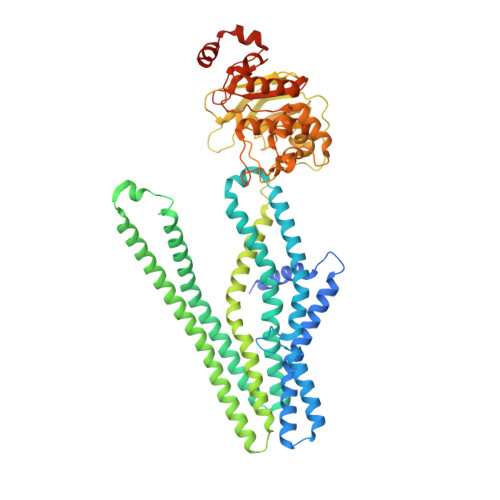
Cryo-EM structure of cadmium-bound human ABCB6.

Choi, S.H.Lee, S.S.Lee, H.Y.Kim, S.Kim, J.W.Jin, M.S.

(2024) Commun Biol 7: 672-672

  • DOI: https://doi.org/10.1038/s42003-024-06377-1
  • Primary Citation Related Structures: 
    8YR3, 8YR4

  • PubMed Abstract: 

    ATP-binding cassette transporter B6 (ABCB6), a protein essential for heme biosynthesis in mitochondria, also functions as a heavy metal efflux pump. Here, we present cryo-electron microscopy structures of human ABCB6 bound to a cadmium Cd(II) ion in the presence of antioxidant thiol peptides glutathione (GSH) and phytochelatin 2 (PC2) at resolutions of 3.2 and 3.1 Å, respectively. The overall folding of the two structures resembles the inward-facing apo state but with less separation between the two halves of the transporter. Two GSH molecules are symmetrically bound to the Cd(II) ion in a bent conformation, with the central cysteine protruding towards the metal. The N-terminal glutamate and C-terminal glycine of GSH do not directly interact with Cd(II) but contribute to neutralizing positive charges of the binding cavity by forming hydrogen bonds and van der Waals interactions with nearby residues. In the presence of PC2, Cd(II) binding to ABCB6 is similar to that observed with GSH, except that two cysteine residues of each PC2 molecule participate in Cd(II) coordination to form a tetrathiolate. Structural comparison of human ABCB6 and its homologous Atm-type transporters indicate that their distinct substrate specificity might be attributed to variations in the capping residues situated at the top of the substrate-binding cavity.


  • Organizational Affiliation
    • School of Life Sciences, GIST, 123 Cheomdangwagi-ro, Buk-gu, Gwangju, Republic of Korea.

Macromolecule Content 

  • Total Structure Weight: 144.17 kDa 
  • Atom Count: 9,319 
  • Modeled Residue Count: 1,174 
  • Deposited Residue Count: 1,274 
  • Unique protein chains: 1

Macromolecules

Find similar proteins by:|  3D Structure
Entity ID: 1
MoleculeChains  Sequence LengthOrganismDetailsImage
ATP-binding cassette sub-family B member 6
A, B
637Homo sapiensMutation(s): 0 
Gene Names: ABCB6MTABC3PRPUMAT
EC: 7.6.2.5
Membrane Entity: Yes 
UniProt & NIH Common Fund Data Resources
Find proteins for Q9NP58 (Homo sapiens)
Explore Q9NP58 
Go to UniProtKB:  Q9NP58
PHAROS:  Q9NP58
GTEx:  ENSG00000115657 
Entity Groups
Sequence Clusters30% Identity50% Identity70% Identity90% Identity95% Identity100% Identity
UniProt GroupQ9NP58
Sequence Annotations
Expand
Reference Sequence

Biologically Interesting Molecules (External Reference) 

1 Unique

Experimental Data & Validation

Experimental Data

  • Method: ELECTRON MICROSCOPY
  • Resolution: 3.20 Å
  • Aggregation State: PARTICLE 
  • Reconstruction Method: SINGLE PARTICLE 
EM Software:
TaskSoftware PackageVersion
MODEL REFINEMENTPHENIX1.20.1_4487:

Structure Validation

View Full Validation Report


Entry History 

& Funding Information

Deposition Data


Funding OrganizationLocationGrant Number
National Research Foundation (NRF, Korea)Korea, Republic OfNRF-2019M3E5D6063908
National Research Foundation (NRF, Korea)Korea, Republic OfNRF-2021M3A9I4022846
National Research Foundation (NRF, Korea)Korea, Republic OfNRF-2022R1A2C1091278

Revision History  (Full details and data files)

  • Version 1.0: 2024-06-19
    Type: Initial release