Molecular insights into the catalytic mechanism of a phthalate ester hydrolase.
Wang, N., Zhang, N., Sun, M.L., Sun, Y., Dong, Q.Y., Wang, Y., Gu, Z.T., Ding, H.T., Qin, Q.L., Jiang, Y., Chen, X.L., Zhang, Y.Z., Gao, C., Li, C.Y.(2024) J Hazard Mater 476: 135191-135191
- PubMed: 39013318 Search on PubMed
- DOI: https://doi.org/10.1016/j.jhazmat.2024.135191
- Primary Citation Related Structures:
8YQJ, 8YQP - PubMed Abstract:
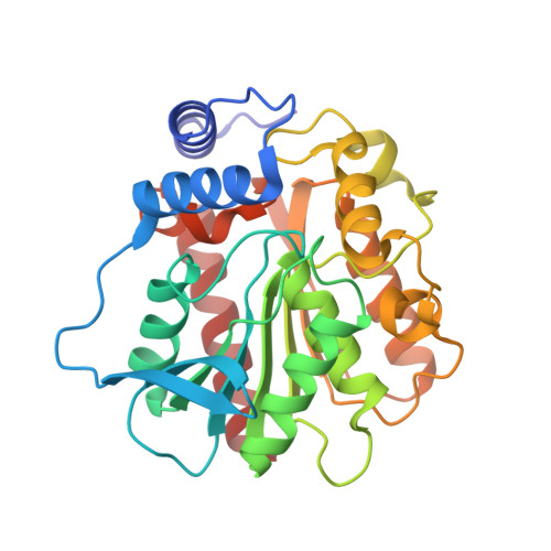
Phthalate esters (PAEs) are emerging hazardous and toxic chemicals that are extensively used as plasticizers or additives. Diethyl phthalate (DEP) and dimethyl phthalate (DMP), two kinds of PAEs, have been listed as the priority pollutants by many countries. PAE hydrolases are the most effective enzymes in PAE degradation, among which family IV esterases are predominate. However, only a few PAE hydrolases have been characterized, and as far as we know, no crystal structure of any PAE hydrolases of the family IV esterases is available to date. HylD1 is a PAE hydrolase of the family IV esterases, which can degrade DMP and DEP. Here, the recombinant HylD1 was characterized. HylD1 maintained a dimer in solution, and functioned under a relatively wide pH range. The crystal structures of HylD1 and its complex with monoethyl phthalate were solved. Residues involved in substrate binding were identified. The catalytic mechanism of HylD1 mediated by the catalytic triad Ser140-Asp231-His261 was further proposed. The hylD1 gene is widely distributed in different environments, suggesting its important role in PAEs degradation. This study provides a better understanding of PAEs hydrolysis, and lays out favorable bases for the rational design of highly-efficient PAEs degradation enzymes for industrial applications in future.
- MOE Key Laboratory of Evolution and Marine Biodiversity, Frontiers Science Center for Deep Ocean Multispheres and Earth System & College of Marine Life Sciences, Ocean University of China, Qingdao, China; State Key Laboratory of Microbial Technology, Marine Biotechnology Research Center, Shandong University, Qingdao, China; Joint Research Center for Marine Microbial Science and Technology, Shandong University and Ocean University of China, Qingdao, China.
Organizational Affiliation: