8YP5 | pdb_00008yp5

The structure of MAP2K4 complexed with 5Z7-oxozeaenol


Experimental Data Snapshot

  • Method: X-RAY DIFFRACTION
  • Resolution: 2.50 Å
  • R-Value Free: 
    0.326 (Depositor), 0.335 (DCC) 
  • R-Value Work: 
    0.233 (Depositor), 0.252 (DCC) 
  • R-Value Observed: 
    0.237 (Depositor) 

Starting Model: experimental
View more details

wwPDB Validation 3D Report Full Report

Validation slider image for 8YP5

Ligand Structure Quality Assessment 


This is version 1.0 of the entry. See complete history

Literature

Conserved gatekeeper methionine regulates the binding and access of kinase inhibitors to ATP sites of MAP2K1, 4, and 7: Clues for developing selective inhibitors.

Yumura, S.Kitagawa, D.Moritsugu, K.Nakayama, A.Shinada, T.Sawa, M.Kinoshita, T.

(2024) Bioorg Med Chem Lett 112: 129914-129914

  • DOI: https://doi.org/10.1016/j.bmcl.2024.129914
  • Primary Citation Related Structures: 
    8YP4, 8YP5

  • PubMed Abstract: 

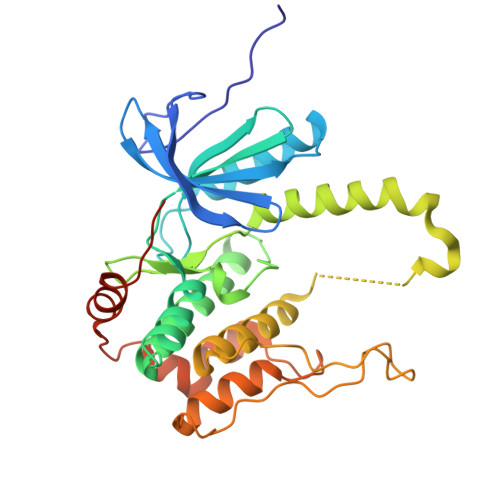
    Mitogen-activated protein kinase kinases (MAP2Ks) 1, 4, and 7 are potential targets for treating various diseases. Here, we solved the crystal structures of MAP2K1 and MAP2K4 complexed with covalent inhibitor 5Z-7-oxozeaenol (5Z7O). The elucidated structures showed that 5Z7O was non-covalently bound to the ATP binding site of MAP2K4, while it covalently attached to cysteine at the DFG-1 position of the deep ATP site of MAP2K1. In contrast, we previously showed that 5Z7O covalently binds to MAP2K7 via another cysteine on the solvent-accessible edge of the ATP site. Structural analyses and molecular dynamics calculations indicated that the configuration and mobility of conserved gatekeeper methionine located at the central ATP site regulated the binding and access of 5Z7O to the ATP site of MAP2Ks. These structural features provide clues for developing highly potent and selective inhibitors against MAP2Ks. Abbreviations: ATP, adenosine triphosphate; FDA, Food and Drug Administration; MAP2Ks, mitogen-activated protein kinase kinases; MD, molecular dynamics; NSCLC, non-small cell lung cancer; 5Z7O, 5Z-7-oxozeaenol; PDB, protein data bank; RMSD, root-mean-square deviation.


  • Organizational Affiliation
    • Graduate School of Science, Osaka Metropolitan University, 1-2 Gakuen-cho, Naka-ku, Sakai, Osaka 599-8570, Japan.

Macromolecule Content 

  • Total Structure Weight: 37.86 kDa 
  • Atom Count: 2,574 
  • Modeled Residue Count: 307 
  • Deposited Residue Count: 327 
  • Unique protein chains: 1

Macromolecules

Find similar proteins by:|  3D Structure
Entity ID: 1
MoleculeChains  Sequence LengthOrganismDetailsImage
Dual specificity mitogen-activated protein kinase kinase 4327Homo sapiensMutation(s): 0 
Gene Names: MAP2K4JNKK1MEK4MKK4PRKMK4SEK1SERK1SKK1
EC: 2.7.12.2
UniProt & NIH Common Fund Data Resources
Find proteins for P45985 (Homo sapiens)
Explore P45985 
Go to UniProtKB:  P45985
PHAROS:  P45985
GTEx:  ENSG00000065559 
Entity Groups
Sequence Clusters30% Identity50% Identity70% Identity90% Identity95% Identity100% Identity
UniProt GroupP45985
Sequence Annotations
Expand
Reference Sequence

Small Molecules

Ligands 2 Unique
IDChains Name / Formula / InChI Key2D Diagram3D Interactions
1FM
(Subject of Investigation/LOI)

Query on 1FM


Download:Ideal Coordinates CCD File
B [auth A](3S,5Z,8S,9S,11E)-8,9,16-trihydroxy-14-methoxy-3-methyl-3,4,9,10-tetrahydro-1H-2-benzoxacyclotetradecine-1,7(8H)-dione
C19 H22 O7
NEQZWEXWOFPKOT-BYRRXHGESA-N
MG
(Subject of Investigation/LOI)

Query on MG


Download:Ideal Coordinates CCD File
C [auth A]MAGNESIUM ION
Mg
JLVVSXFLKOJNIY-UHFFFAOYSA-N

Experimental Data & Validation

Experimental Data

  • Method: X-RAY DIFFRACTION
  • Resolution: 2.50 Å
  • R-Value Free:  0.326 (Depositor), 0.335 (DCC) 
  • R-Value Work:  0.233 (Depositor), 0.252 (DCC) 
  • R-Value Observed: 0.237 (Depositor) 
Space Group: C 2 2 21
Unit Cell:
Length ( Å )Angle ( ˚ )
a = 68.81α = 90
b = 118.032β = 90
c = 81.52γ = 90
Software Package:
Software NamePurpose
PHENIXrefinement
XDSdata reduction
XDSdata scaling
PHASERphasing

Structure Validation

View Full Validation Report


Ligand Structure Quality Assessment 


Entry History 

& Funding Information

Deposition Data


Funding OrganizationLocationGrant Number
Not funded--

Revision History  (Full details and data files)

  • Version 1.0: 2025-01-22
    Type: Initial release