8YH5 | pdb_00008yh5

A3R-Gi complex bound to i6A


Experimental Data Snapshot

  • Method: ELECTRON MICROSCOPY
  • Resolution: 3.66 Å
  • Aggregation State: PARTICLE 
  • Reconstruction Method: SINGLE PARTICLE 

wwPDB Validation 3D Report Full Report

Validation slider image for 8YH5

This is version 1.2 of the entry. See complete history

Literature

Structural insights into the agonist selectivity of the adenosine A 3 receptor.

Oshima, H.S.Ogawa, A.Sano, F.K.Akasaka, H.Kawakami, T.Iwama, A.Okamoto, H.H.Nagiri, C.Wei, F.Y.Shihoya, W.Nureki, O.

(2024) Nat Commun 15: 9294-9294

  • DOI: https://doi.org/10.1038/s41467-024-53473-1
  • Primary Citation Related Structures: 
    8YH0, 8YH2, 8YH3, 8YH5, 8YH6

  • PubMed Abstract: 

    Adenosine receptors play pivotal roles in physiological processes. Adenosine A 3 receptor (A 3 R), the most recently identified adenosine receptor, is expressed in various tissues, exhibiting important roles in neuron, heart, and immune cells, and is often overexpressed in tumors, highlighting the therapeutic potential of A 3 R-selective agents. Recently, we identified RNA-derived N 6 -methyladenosine (m 6 A) as an endogenous agonist for A 3 R, suggesting the relationship between RNA-derived modified adenosine and A 3 R. Despite extensive studies on the other adenosine receptors, the selectivity mechanism of A 3 R, especially for A 3 R-selective agonists such as m 6 A and namodenoson, remained elusive. Here, we identify tRNA-derived N 6 -isopentenyl adenosine (i 6 A) as an A 3 R-selective ligand via screening of modified nucleosides against the adenosine receptors. Like m 6 A, i 6 A is found in the human body and may be an endogenous A 3 R ligand. Our cryo-EM analyses elucidate the A 3 R-G i complexes bound to adenosine, 5'-N-ethylcarboxamidoadenosine (NECA), m 6 A, i 6 A, and namodenoson at overall resolutions of 3.27 Å (adenosine), 2.86 Å (NECA), 3.19 Å (m 6 A), 3.28 Å (i 6 A), and 3.20 Å (namodenoson), suggesting the selectivity and activation mechanism of A 3 R. We further conduct structure-guided engineering of m 6 A-insensitive A 3 R, which may aid future research targeting m 6 A and A 3 R, providing a molecular basis for future drug discovery.


  • Organizational Affiliation
    • Department of Biological Sciences, Graduate School of Science, The University of Tokyo, Tokyo, Japan.

Macromolecule Content 

  • Total Structure Weight: 254.65 kDa 
  • Atom Count: 8,695 
  • Modeled Residue Count: 1,103 
  • Deposited Residue Count: 2,295 
  • Unique protein chains: 4

Macromolecules

Find similar proteins by:|  3D Structure
Entity ID: 1
MoleculeChains  Sequence LengthOrganismDetailsImage
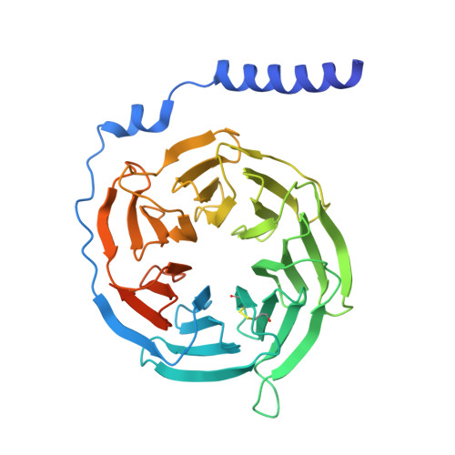
Guanine nucleotide-binding protein G(I)/G(S)/G(T) subunit beta-1A [auth B]375Rattus rattusMutation(s): 0 
Gene Names: GNB1
UniProt
Find proteins for P54311 (Rattus norvegicus)
Explore P54311 
Go to UniProtKB:  P54311
Entity Groups
Sequence Clusters30% Identity50% Identity70% Identity90% Identity95% Identity100% Identity
UniProt GroupP54311
Sequence Annotations
Expand
Reference Sequence
Find similar proteins by:|  3D Structure
Entity ID: 2
MoleculeChains  Sequence LengthOrganismDetailsImage
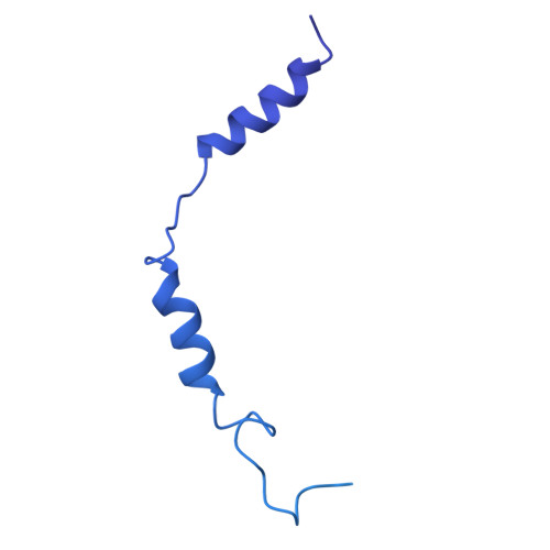
Guanine nucleotide-binding protein G(I)/G(S)/G(O) subunit gamma-2,Guanine nucleotide-binding protein G(i) subunit alpha-1 chimeraB [auth G],
D [auth A]
433Homo sapiensBos taurus
This entity is chimeric
Mutation(s): 0 
Gene Names: GNG2GNAI1
EC: 3.6.5
Membrane Entity: Yes 
UniProt & NIH Common Fund Data Resources
Find proteins for P63096 (Homo sapiens)
Explore P63096 
Go to UniProtKB:  P63096
PHAROS:  P63096
GTEx:  ENSG00000127955 
Entity Groups
Sequence Clusters30% Identity50% Identity70% Identity90% Identity95% Identity100% Identity
UniProt GroupP63096
Sequence Annotations
Expand
Reference Sequence
Find similar proteins by:|  3D Structure
Entity ID: 3
MoleculeChains  Sequence LengthOrganismDetailsImage
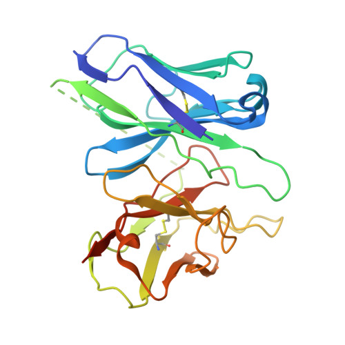
scfv16C [auth S]260Mus musculusMutation(s): 0 
Membrane Entity: Yes 
UniProt
Find proteins for A0A0B4J1H6 (Mus musculus)
Explore A0A0B4J1H6 
Go to UniProtKB:  A0A0B4J1H6
Entity Groups
Sequence Clusters30% Identity50% Identity70% Identity90% Identity95% Identity100% Identity
UniProt GroupA0A0B4J1H6
Sequence Annotations
Expand
Reference Sequence
Find similar proteins by:|  3D Structure
Entity ID: 4
MoleculeChains  Sequence LengthOrganismDetailsImage
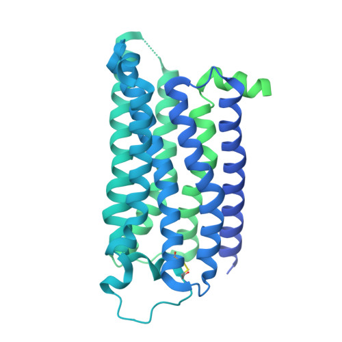
Hemagglutinin,Adenosine receptor A3,GFP chimeraE [auth R]794Influenza A virus (A/Victoria/3/1975(H3N2))Ovis arieshuman respiratory syncytial virusMutation(s): 0 
Gene Names: HAADORA3
Membrane Entity: Yes 
UniProt
Find proteins for P35342 (Ovis aries)
Explore P35342 
Go to UniProtKB:  P35342
Entity Groups
Sequence Clusters30% Identity50% Identity70% Identity90% Identity95% Identity100% Identity
UniProt GroupP35342
Sequence Annotations
Expand
Reference Sequence

Small Molecules

Ligands 1 Unique
IDChains Name / Formula / InChI Key2D Diagram3D Interactions
ZIR
(Subject of Investigation/LOI)

Query on ZIR


Download:Ideal Coordinates CCD File
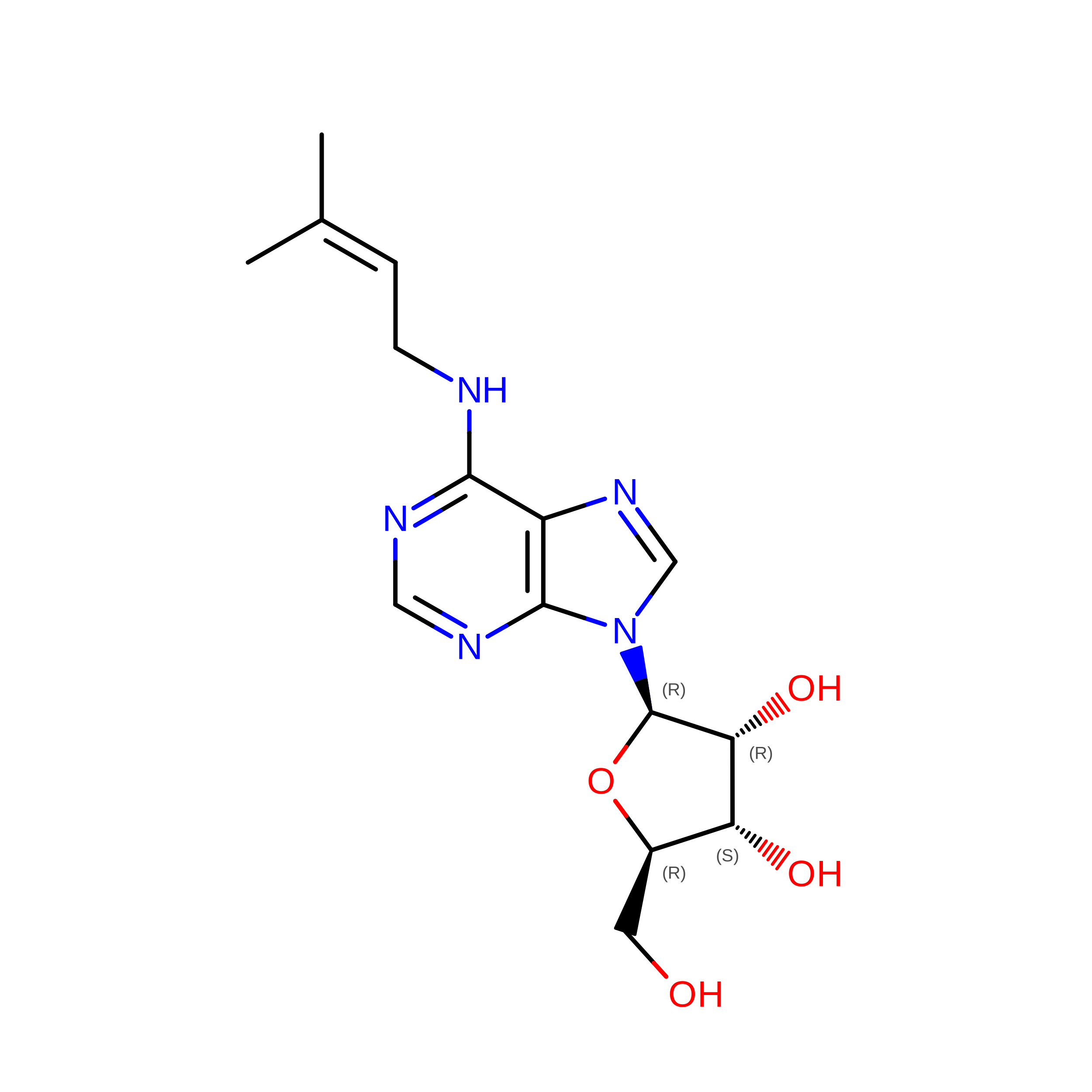
F [auth R]N-(3-methylbut-2-en-1-yl)adenosine
C15 H21 N5 O4
USVMJSALORZVDV-SDBHATRESA-N

Experimental Data & Validation

Experimental Data

  • Method: ELECTRON MICROSCOPY
  • Resolution: 3.66 Å
  • Aggregation State: PARTICLE 
  • Reconstruction Method: SINGLE PARTICLE 
EM Software:
TaskSoftware PackageVersion
MODEL REFINEMENTPHENIX

Structure Validation

View Full Validation Report


Entry History 

& Funding Information

Deposition Data


Funding OrganizationLocationGrant Number
Japan Society for the Promotion of Science (JSPS)Japan21H05037

Revision History  (Full details and data files)

  • Version 1.0: 2024-11-06
    Type: Initial release
  • Version 1.1: 2024-11-27
    Changes: Data collection, Database references
  • Version 1.2: 2025-06-18
    Changes: Data collection