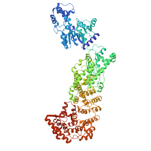
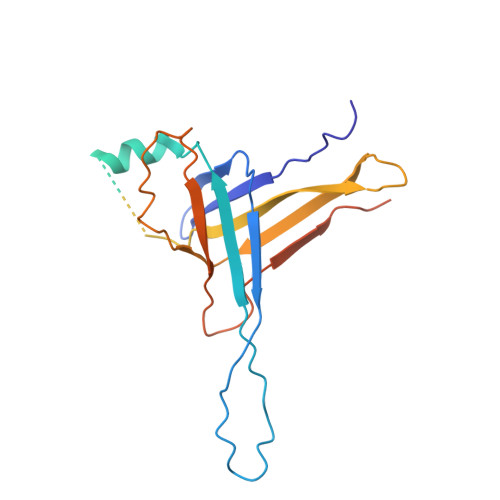
The NADase activity of the defense-associated sirtuins (DSRs) is activated by the phage tail tube protein (TTP). Herein, we report cryo-EM structures of a free-state Bacillus subtilis DSR2 tetramer and a fragment of the tetramer, a phage SPR tail tube, and two DSR2-TTP complexes. DSR2 contains an N-terminal SIR2 domain, a middle domain (MID) and a C-terminal domain (CTD). The DSR2 CTD harbors the α-solenoid tandem-repeats like the HEAT-repeat proteins. DSR2 assembles into a tetramer with four SIR2 clustered at the center, and two intertwined MID-CTD chains flank the SIR2 core. SPR TTPs self-assemble into a tube-like complex. Upon DSR2 binding, the D1 domain of SPR TTP is captured between the HEAT-repeats domains of DSR2, which conflicts with TTPs self-assembly. Binding of TTPs induces conformational changes in DSR2 tetramer, resulting in increase of the NAD + pocket volume in SIR2, thus activates the NADase activity and leads to cellular NAD + depletion.
Organizational Affiliation:
NHC Key Laboratory of Systems Biology of Pathogens, National Institute of Pathogen Biology, Chinese Academy of Medical Sciences & Peking Union Medical College, Beijing, China.
Key Laboratory of Pathogen Infection Prevention and Control (Ministry of Education), National Institute of Pathogen Biology, Chinese Academy of Medical Sciences & Peking Union Medical College, Beijing, China.
State Key Laboratory of Respiratory Health and Multimorbidity, National Institute of Pathogen Biology, Chinese Academy of Medical Sciences & Peking Union Medical College, Beijing, China.
Yanan medical college of Yanan university, Yanan, China.
Beijing National Laboratory for Condensed Matter Physics, Institute of Physics, Chinese Academy of Sciences, Beijing, China.
National Clinical Laboratory on Tuberculosis, Beijing Key Laboratory for Drug-resistant Tuberculosis Research Beijing Chest Hospital, Capital Medical University, Beijing Tuberculosis and Thoracic Tumor Institute, Beijing, China.
Beijing Advanced Innovation Center for Structural Biology, Tsinghua-Peking Joint Center for Life Sciences, School of Life Sciences, Tsinghua University, Beijing, China.
NHC Key Laboratory of Systems Biology of Pathogens, National Institute of Pathogen Biology, Chinese Academy of Medical Sciences & Peking Union Medical College, Beijing, China. gaoxiaopan@pumc.edu.cn.
Key Laboratory of Pathogen Infection Prevention and Control (Ministry of Education), National Institute of Pathogen Biology, Chinese Academy of Medical Sciences & Peking Union Medical College, Beijing, China. gaoxiaopan@pumc.edu.cn.
State Key Laboratory of Respiratory Health and Multimorbidity, National Institute of Pathogen Biology, Chinese Academy of Medical Sciences & Peking Union Medical College, Beijing, China. gaoxiaopan@pumc.edu.cn.
Beijing National Laboratory for Condensed Matter Physics, Institute of Physics, Chinese Academy of Sciences, Beijing, China. hongtao.zhu@iphy.ac.cn.
University of Chinese Academy of Sciences, Beijing, China. hongtao.zhu@iphy.ac.cn.
Songshan Lake Materials Laboratory, Dongguan, China. hongtao.zhu@iphy.ac.cn.
NHC Key Laboratory of Systems Biology of Pathogens, National Institute of Pathogen Biology, Chinese Academy of Medical Sciences & Peking Union Medical College, Beijing, China. cui.sheng@ipb.pumc.edu.cn.
Key Laboratory of Pathogen Infection Prevention and Control (Ministry of Education), National Institute of Pathogen Biology, Chinese Academy of Medical Sciences & Peking Union Medical College, Beijing, China. cui.sheng@ipb.pumc.edu.cn.
State Key Laboratory of Respiratory Health and Multimorbidity, National Institute of Pathogen Biology, Chinese Academy of Medical Sciences & Peking Union Medical College, Beijing, China. cui.sheng@ipb.pumc.edu.cn.