Lowering pH optimum of activity of SshEstI, a slightly alkaliphilic archaeal esterase of the hormone-sensitive lipase family.
Ohara, K., Oshima, Y., Unno, H., Nagano, S., Kusunoki, M., Takahashi, S., Waki, T., Yamashita, S., Nakayama, T.(2024) J Biosci Bioeng 138: 188-195
- PubMed: 38918133 Search on PubMed
- DOI: https://doi.org/10.1016/j.jbiosc.2024.05.010
- Primary Citation Related Structures:
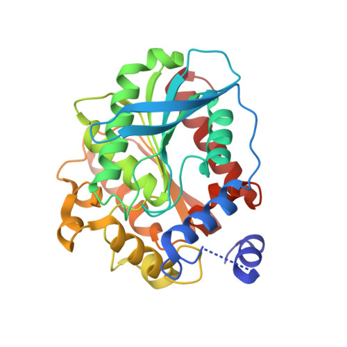
8YFY, 8YFZ - PubMed Abstract:
SshEstI, a carboxylesterase from the thermoacidophilic archaeon Saccharolobus shibatae, is a member of the hormone-sensitive lipase family that displays slightly alkaliphilic activity with an optimum activity at pH 8.0. In this study, three distinct strategies were explored to confer acidophilic properties to SshEstI. The first strategy involved engineering the oxyanion hole by replacing Gly81 with serine or aspartic acid. The G81S mutant showed optimum activity at pH 7.0, whereas the aspartic acid mutant (G81D) rendered the enzyme slightly acidophilic with optimum activity observed at pH 6.0; however, k cat and k cat /K m values were reduced by these substitutions. The second strategy involved examining the effects of surfactant additives on the pH-activity profiles of SshEstI. The results showed that cetyltrimethylammonium bromide (CTAB) enhanced wild-type enzyme (WT) activity at acidic pH values. In the presence of 0.1 mM CTAB, G81S and G81D were acidophilic enzymes with optimum activity at pH 6.0 and 4.0, respectively, although their enzyme activities were low. The third strategy involved engineering the active site to resemble that of kumamolisin-As (kuma-As), an acidophilic peptidase of the sedolisin family. The catalytic triad of kuma-As was exchanged into SshEstI using site-directed mutagenesis. X-ray crystallographic analysis of the mutants (H274D and H274E) revealed that the potential hydrogen donor-acceptor distances around the active site of WT were fully maintained in these mutants. However, these mutants were inactive at pH 4-8.
- Department of Biomolecular Engineering, Graduate School of Engineering, Tohoku University, Sendai, Miyagi 980-8579, Japan.
Organizational Affiliation: