


Crystal structure of human Cu-Zn Superoxide Dismutase 1 in complex with 1,2,10-Decanetriol
Aouti, S., Padmanabhan, B.To be published.
Experimental Data Snapshot
Starting Model: experimental
View more details
Entity ID: 1 | |||||
|---|---|---|---|---|---|
| Molecule | Chains | Sequence Length | Organism | Details | Image |
| Superoxide dismutase [Cu-Zn] | 154 | Homo sapiens | Mutation(s): 0 Gene Names: SOD1 EC: 1.15.1.1 (PDB Primary Data), 1.8 (UniProt) | 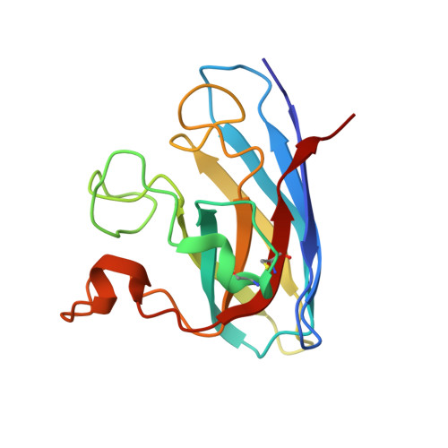 | |
UniProt & NIH Common Fund Data Resources | |||||
PHAROS: P00441 GTEx: ENSG00000142168 | |||||
Entity Groups | |||||
| Sequence Clusters | 30% Identity50% Identity70% Identity90% Identity95% Identity100% Identity | ||||
| UniProt Group | P00441 | ||||
Sequence AnnotationsExpand | |||||
Reference Sequence | |||||
| Ligands 3 Unique | |||||
|---|---|---|---|---|---|
| ID | Chains | Name / Formula / InChI Key | 2D Diagram | 3D Interactions | |
| A1LYN (Subject of Investigation/LOI) Download:Ideal Coordinates CCD File | BA [auth H] DA [auth I] FA [auth J] GA [auth J] M [auth A] | (2S)-decane-1,2,10-triol C10 H22 O3 RHINSRUDDXGHLV-JTQLQIEISA-N |  | ||
| GOL Download:Ideal Coordinates CCD File | L [auth A], P [auth B], S [auth C], V [auth D], Y [auth F] | GLYCEROL C3 H8 O3 PEDCQBHIVMGVHV-UHFFFAOYSA-N |  | ||
| ZN Download:Ideal Coordinates CCD File | AA [auth H] CA [auth I] EA [auth J] K [auth A] N [auth B] | ZINC ION Zn PTFCDOFLOPIGGS-UHFFFAOYSA-N |  | ||
| Length ( Å ) | Angle ( ˚ ) |
|---|---|
| a = 163.666 | α = 90 |
| b = 202.311 | β = 90 |
| c = 143.668 | γ = 90 |
| Software Name | Purpose |
|---|---|
| PHENIX | refinement |
| autoPROC | data reduction |
| autoPROC | data scaling |
| PHASER | phasing |
| Funding Organization | Location | Grant Number |
|---|---|---|
| Indian Council of Medical Research | India | -- |


