8YB1 | pdb_00008yb1

XFEL crystal structure of the reduced form of F87A/F393H P450BM3 with N-enanthyl-L-prolyl-L-phenylalanine in complex with styrene


Experimental Data Snapshot

  • Method: X-RAY DIFFRACTION
  • Resolution: 1.60 Å
  • R-Value Free: 
    0.214 (Depositor), 0.214 (DCC) 
  • R-Value Work: 
    0.184 (Depositor), 0.184 (DCC) 
  • R-Value Observed: 
    0.185 (Depositor) 

Starting Model: experimental
View more details

wwPDB Validation 3D Report Full Report

Validation slider image for 8YB1

Ligand Structure Quality Assessment 


This is version 1.2 of the entry. See complete history

Literature

XFEL crystallography reveals catalytic cycle dynamics during non-native substrate oxidation by cytochrome P450BM3.

Nagao, S.Kuwano, W.Tosha, T.Yamashita, K.Stanfield, J.K.Kasai, C.Ariyasu, S.Hirata, K.Ueno, G.Murakami, H.Ago, H.Yamamoto, M.Shoji, O.Sugimoto, H.Kubo, M.

(2025) Commun Chem 8: 63-63

  • DOI: https://doi.org/10.1038/s42004-025-01440-2
  • Primary Citation Related Structures: 
    8YAY, 8YAZ, 8YB0, 8YB1, 8YB2, 8YB3, 9KT5

  • PubMed Abstract: 

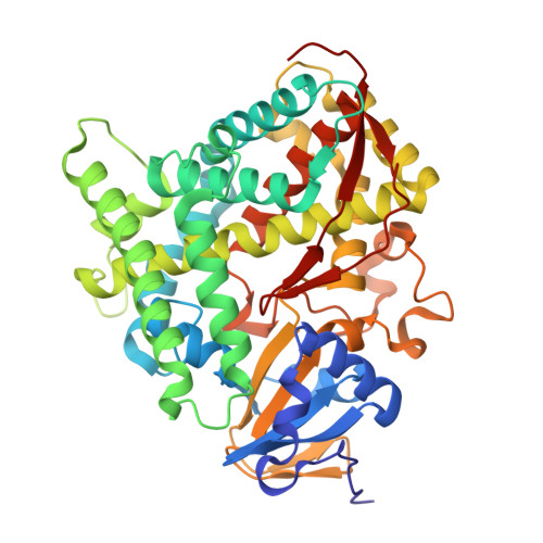
    Cytochrome P450s are haem-containing enzymes, catalysing the regio- and stereospecific oxidation of non-activated hydrocarbons. Among these, the bacterial P450BM3 is a promising biocatalyst due to its high enzymatic activity. Given the significant conformational flexibility of this enzyme, understanding protein-substrate interactions and associated structural dynamics are crucial for designing P450BM3-based biocatalysts. Herein, employing an X-ray free electron laser in combination with freeze-trap crystallography and spectroscopy techniques, we captured the intact structures of engineered P450BM3s in the initial stages of catalysis during styrene epoxidation, in the presence of a decoy molecule. We found that the iron reduction significantly altered the active-site orientation of styrene, driven by structural changes in surrounding helices and hydrogen-bonding networks. Oxygen binding to iron further stabilised its productive orientation, providing a molecular basis for the experimentally observed enzyme kinetics and enantioselectivities. This study reveals the substrate dynamics of a P450 enzyme, showcasing how changes in haem chemistry affect enzyme structure and substrate orientation.


  • Organizational Affiliation
    • Graduate School of Science, University of Hyogo, Ako-gun, Hyogo, Japan.

Macromolecule Content 

  • Total Structure Weight: 106.61 kDa 
  • Atom Count: 8,321 
  • Modeled Residue Count: 910 
  • Deposited Residue Count: 910 
  • Unique protein chains: 1

Macromolecules

Find similar proteins by:|  3D Structure
Entity ID: 1
MoleculeChains  Sequence LengthOrganismDetailsImage
Bifunctional cytochrome P450/NADPH--P450 reductase
A, B
455Priestia megaterium NBRC 15308 = ATCC 14581Mutation(s): 2 
EC: 1.14.14.1 (PDB Primary Data), 1.6.2.4 (PDB Primary Data)
UniProt
Find proteins for P14779 (Priestia megaterium (strain ATCC 14581 / DSM 32 / CCUG 1817 / JCM 2506 / NBRC 15308 / NCIMB 9376 / NCTC 10342 / NRRL B-14308 / VKM B-512 / Ford 19))
Explore P14779 
Go to UniProtKB:  P14779
Entity Groups
Sequence Clusters30% Identity50% Identity70% Identity90% Identity95% Identity100% Identity
UniProt GroupP14779
Sequence Annotations
Expand
Reference Sequence

Small Molecules

Ligands 4 Unique
IDChains Name / Formula / InChI Key2D Diagram3D Interactions
HEM
(Subject of Investigation/LOI)

Query on HEM


Download:Ideal Coordinates CCD File
C [auth A],
F [auth B]
PROTOPORPHYRIN IX CONTAINING FE
C34 H32 Fe N4 O4
KABFMIBPWCXCRK-RGGAHWMASA-L
D0L

Query on D0L


Download:Ideal Coordinates CCD File
D [auth A],
G [auth B]
(2S)-2-[[(2S)-1-heptylpyrrolidin-2-yl]carbonylamino]-3-phenyl-propanoic acid
C21 H32 N2 O3
UZXMNGCHFSENBI-OALUTQOASA-N
SYN
(Subject of Investigation/LOI)

Query on SYN


Download:Ideal Coordinates CCD File
E [auth A],
H [auth B]
ethenylbenzene
C8 H8
PPBRXRYQALVLMV-UHFFFAOYSA-N
GOL

Query on GOL


Download:Ideal Coordinates CCD File
I [auth B],
J [auth B],
K [auth B]
GLYCEROL
C3 H8 O3
PEDCQBHIVMGVHV-UHFFFAOYSA-N

Experimental Data & Validation

Experimental Data

  • Method: X-RAY DIFFRACTION
  • Resolution: 1.60 Å
  • R-Value Free:  0.214 (Depositor), 0.214 (DCC) 
  • R-Value Work:  0.184 (Depositor), 0.184 (DCC) 
  • R-Value Observed: 0.185 (Depositor) 
Space Group: P 21 21 21
Unit Cell:
Length ( Å )Angle ( ˚ )
a = 58.86α = 90
b = 128.49β = 90
c = 149.51γ = 90
Software Package:
Software NamePurpose
PHENIXrefinement
CrystFELdata reduction
CrystFELdata scaling
PHENIXphasing

Structure Validation

View Full Validation Report


Ligand Structure Quality Assessment 


Entry History 

& Funding Information

Deposition Data


Funding OrganizationLocationGrant Number
Japan Science and TechnologyJapanJPMJCR15P3
Japan Society for the Promotion of Science (JSPS)JapanJP19H05784
Japan Society for the Promotion of Science (JSPS)JapanJP18KK0397
Japan Society for the Promotion of Science (JSPS)JapanJP19K22403

Revision History  (Full details and data files)

  • Version 1.0: 2025-02-12
    Type: Initial release
  • Version 1.1: 2025-03-26
    Changes: Database references
  • Version 1.2: 2025-04-02
    Changes: Database references