8Y9B | pdb_00008y9b

TcdB1 in complex with mini-binder


Experimental Data Snapshot

  • Method: ELECTRON MICROSCOPY
  • Resolution: 3.20 Å
  • Aggregation State: PARTICLE 
  • Reconstruction Method: SINGLE PARTICLE 

Starting Models: in silico, experimental
View more details

wwPDB Validation 3D Report Full Report

Validation slider image for 8Y9B

This is version 1.2 of the entry. See complete history

Literature

De novo design of mini-protein binders broadly neutralizing Clostridioides difficile toxin B variants.

Lv, X.Zhang, Y.Sun, K.Yang, Q.Luo, J.Tao, L.Lu, P.

(2024) Nat Commun 15: 8521-8521

  • DOI: https://doi.org/10.1038/s41467-024-52582-1
  • Primary Citation Related Structures: 
    8Y9B, 8Y9C

  • PubMed Abstract: 

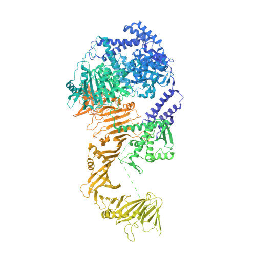
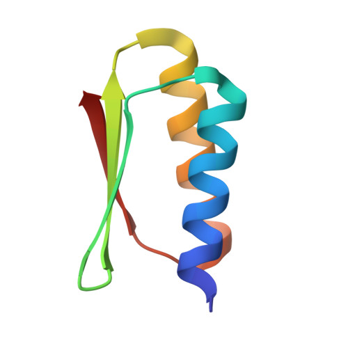
    Clostridioides difficile toxin B (TcdB) is the key virulence factor accounting for C. difficile infection-associated symptoms. Effectively neutralizing different TcdB variants with a universal solution poses a significant challenge. Here we present the de novo design and characterization of pan-specific mini-protein binders against major TcdB subtypes. Our design successfully binds to the first receptor binding interface (RBI-1) of the varied TcdB subtypes, exhibiting affinities ranging from 20 pM to 10 nM. The cryo-electron microscopy (cryo-EM) structures of the mini protein binder in complex with TcdB1 and TcdB4 are consistent with the computational design models. The engineered and evolved variants of the mini-protein binder and chondroitin sulfate proteoglycan 4 (CSPG4), another natural receptor that binds to the second RBI (RBI-2) of TcdB, better neutralize major TcdB variants both in cells and in vivo, as demonstrated by the colon-loop assay using female mice. Our findings provide valuable starting points for the development of therapeutics targeting C. difficile infections (CDI).


  • Organizational Affiliation
    • Research Center for Industries of the Future, Westlake University, Hangzhou, Zhejiang, 310024, China.

Macromolecule Content 

  • Total Structure Weight: 278.27 kDa 
  • Atom Count: 13,380 
  • Modeled Residue Count: 1,672 
  • Deposited Residue Count: 2,437 
  • Unique protein chains: 2

Macromolecules

Find similar proteins by:|  3D Structure
Entity ID: 1
MoleculeChains  Sequence LengthOrganismDetailsImage
Toxin BA [auth B]2,372Clostridioides difficileMutation(s): 0 
Gene Names: tcdBtoxB
EC: 3.4.22
UniProt
Find proteins for P18177 (Clostridioides difficile)
Explore P18177 
Go to UniProtKB:  P18177
Entity Groups
Sequence Clusters30% Identity50% Identity70% Identity90% Identity95% Identity100% Identity
UniProt GroupP18177
Sequence Annotations
Expand
Reference Sequence
Find similar proteins by:|  3D Structure
Entity ID: 2
MoleculeChains  Sequence LengthOrganismDetailsImage
De novo design mini-binderB [auth A]65synthetic constructMutation(s): 0 

Small Molecules

Experimental Data & Validation

Experimental Data

  • Method: ELECTRON MICROSCOPY
  • Resolution: 3.20 Å
  • Aggregation State: PARTICLE 
  • Reconstruction Method: SINGLE PARTICLE 
EM Software:
TaskSoftware PackageVersion
MODEL REFINEMENTPHENIX1.19.2_4158:

Structure Validation

View Full Validation Report


Entry History 

& Funding Information

Deposition Data


Funding OrganizationLocationGrant Number
Ministry of Science and Technology (MoST, China)China2020YFA0909200
National Natural Science Foundation of China (NSFC)China32301219

Revision History  (Full details and data files)

  • Version 1.0: 2024-08-28
    Type: Initial release
  • Version 1.1: 2024-10-09
    Changes: Data collection, Database references, Structure summary
  • Version 1.2: 2024-10-16
    Changes: Data collection, Database references