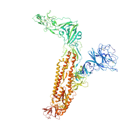
8Y8C

Structure of HCoV-HKU1C spike in the inactive-closed conformation


Experimental Data Snapshot

  • Method: ELECTRON MICROSCOPY
  • Resolution: 2.95 Å
  • Aggregation State: PARTICLE 
  • Reconstruction Method: SINGLE PARTICLE 

wwPDB Validation   3D Report Full Report


This is version 1.0 of the entry. See complete history


Literature

TMPRSS2 and glycan receptors synergistically facilitate coronavirus entry.

Wang, H.Liu, X.Zhang, X.Zhao, Z.Lu, Y.Pu, D.Zhang, Z.Chen, J.Wang, Y.Li, M.Dong, X.Duan, Y.He, Y.Mao, Q.Guo, H.Sun, H.Zhou, Y.Yang, Q.Gao, Y.Yang, X.Cao, H.Guddat, L.Sun, L.Rao, Z.Yang, H.

(2024) Cell 

  • DOI: https://doi.org/10.1016/j.cell.2024.06.016
  • Primary Citation of Related Structures:  
    8Y7X, 8Y7Y, 8Y87, 8Y88, 8Y89, 8Y8A, 8Y8B, 8Y8C, 8Y8D, 8Y8E, 8Y8F, 8Y8G, 8Y8H, 8Y8I, 8Y8J

  • PubMed Abstract: 

    The entry of coronaviruses is initiated by spike recognition of host cellular receptors, involving proteinaceous and/or glycan receptors. Recently, TMPRSS2 was identified as the proteinaceous receptor for HCoV-HKU1 alongside sialoglycan as a glycan receptor. However, the underlying mechanisms for viral entry remain unknown. Here, we investigated the HCoV-HKU1C spike in the inactive, glycan-activated, and functionally anchored states, revealing that sialoglycan binding induces a conformational change of the NTD and promotes the neighboring RBD of the spike to open for TMPRSS2 recognition, exhibiting a synergistic mechanism for the entry of HCoV-HKU1. The RBD of HCoV-HKU1 features an insertion subdomain that recognizes TMPRSS2 through three previously undiscovered interfaces. Furthermore, structural investigation of HCoV-HKU1A in combination with mutagenesis and binding assays confirms a conserved receptor recognition pattern adopted by HCoV-HKU1. These studies advance our understanding of the complex viral-host interactions during entry, laying the groundwork for developing new therapeutics against coronavirus-associated diseases.


  • Organizational Affiliation

    Shanghai Institute for Advanced Immunochemical Studies and School of Life Science and Technology, ShanghaiTech University, Shanghai 201210, China; Shanghai Clinical Research and Trial Center, Shanghai 201210, China.


Macromolecules
Find similar proteins by:  (by identity cutoff)  |  3D Structure
Entity ID: 1
MoleculeChains Sequence LengthOrganismDetailsImage
Spike glycoprotein
A, B, C
1,263Human coronavirus HKU1 (isolate N5)Mutation(s): 10 
Gene Names: S3
UniProt
Find proteins for Q0ZME7 (Human coronavirus HKU1 (isolate N5))
Explore Q0ZME7 
Go to UniProtKB:  Q0ZME7
Entity Groups  
Sequence Clusters30% Identity50% Identity70% Identity90% Identity95% Identity100% Identity
UniProt GroupQ0ZME7
Glycosylation
Glycosylation Sites: 8
Sequence Annotations
Expand
  • Reference Sequence
Small Molecules
Ligands 1 Unique
IDChains Name / Formula / InChI Key2D Diagram3D Interactions
NAG
Query on NAG

Download Ideal Coordinates CCD File 
AA [auth C]
BA [auth C]
D [auth A]
E [auth A]
F [auth A]
AA [auth C],
BA [auth C],
D [auth A],
E [auth A],
F [auth A],
G [auth A],
H [auth A],
I [auth A],
J [auth A],
K [auth A],
L [auth A],
M [auth B],
N [auth B],
O [auth B],
P [auth B],
Q [auth B],
R [auth B],
S [auth B],
T [auth B],
U [auth C],
V [auth C],
W [auth C],
X [auth C],
Y [auth C],
Z [auth C]
2-acetamido-2-deoxy-beta-D-glucopyranose
C8 H15 N O6
OVRNDRQMDRJTHS-FMDGEEDCSA-N
Experimental Data & Validation

Experimental Data

  • Method: ELECTRON MICROSCOPY
  • Resolution: 2.95 Å
  • Aggregation State: PARTICLE 
  • Reconstruction Method: SINGLE PARTICLE 

Structure Validation

View Full Validation Report


Entry History & Funding Information

Deposition Data


Funding OrganizationLocationGrant Number
National Natural Science Foundation of China (NSFC)China92169109

Revision History  (Full details and data files)

  • Version 1.0: 2024-07-17
    Type: Initial release