


Structure of Staphylococcus aureus RecJ
Cheng, K., Wang, Y.To be published.
Experimental Data Snapshot
Starting Model: experimental
View more details
Entity ID: 1 | |||||
|---|---|---|---|---|---|
| Molecule | Chains | Sequence Length | Organism | Details | Image |
| Single-stranded-DNA-specific exonuclease RecJ | 757 | Staphylococcus aureus | Mutation(s): 0 Gene Names: recJ, C7M54_09320, DD547_01719, EP54_09380, EQ90_02295, FAF17_07585, GO736_02485, GO814_13090, GO942_04975, GQX37_04600... | 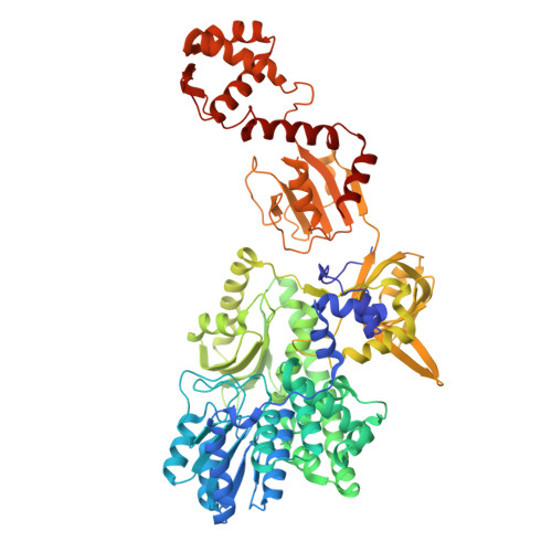 | |
UniProt | |||||
Entity Groups | |||||
| Sequence Clusters | 30% Identity50% Identity70% Identity90% Identity95% Identity100% Identity | ||||
| UniProt Group | Q2FXT9 | ||||
Sequence AnnotationsExpand | |||||
Reference Sequence | |||||
| Ligands 2 Unique | |||||
|---|---|---|---|---|---|
| ID | Chains | Name / Formula / InChI Key | 2D Diagram | 3D Interactions | |
| SO4 (Subject of Investigation/LOI) Download:Ideal Coordinates CCD File | F [auth A] G [auth A] H [auth A] K [auth B] L [auth B] | SULFATE ION O4 S QAOWNCQODCNURD-UHFFFAOYSA-L |  | ||
| MG (Subject of Investigation/LOI) Download:Ideal Coordinates CCD File | D [auth A] E [auth A] I [auth B] J [auth B] N [auth C] | MAGNESIUM ION Mg JLVVSXFLKOJNIY-UHFFFAOYSA-N |  | ||
| Length ( Å ) | Angle ( ˚ ) |
|---|---|
| a = 199.313 | α = 90 |
| b = 199.313 | β = 90 |
| c = 62.764 | γ = 120 |
| Software Name | Purpose |
|---|---|
| PHENIX | refinement |
| autoPROC | data processing |
| autoPROC | data reduction |
| PHASER | phasing |
| Coot | model building |
| Funding Organization | Location | Grant Number |
|---|---|---|
| National Natural Science Foundation of China (NSFC) | China | 32100017 |
| National Natural Science Foundation of China (NSFC) | China | 32270043 |


