Allosteric inhibition of IgE-Fc epsilon RI interactions by simultaneous targeting of IgE F(ab')2 epitopes.
Hirano, T., Koyanagi, A., Ago, H., Yamamoto, M., Kitaura, J., Kasai, M., Okumura, K.(2024) Commun Biol 7: 1042-1042
- PubMed: 39179708 Search on PubMedSearch on PubMed Central
- DOI: https://doi.org/10.1038/s42003-024-06633-4
- Primary Citation Related Structures:
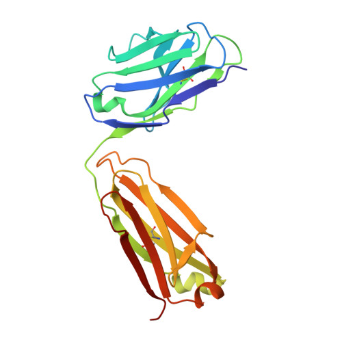
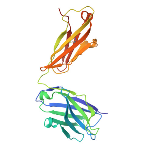
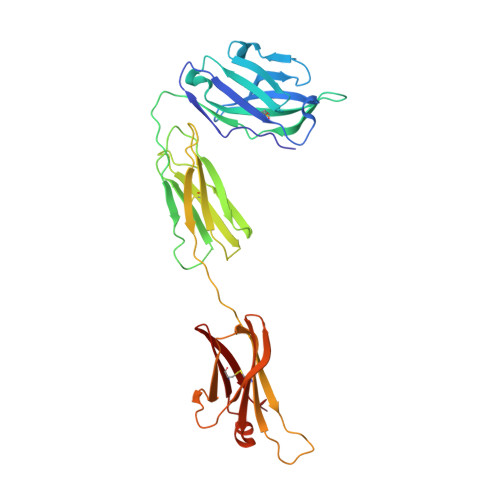
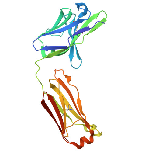
8XX0 - PubMed Abstract:
Immunoglobulin E (IgE) plays pivotal roles in allergic diseases through interaction with a high-affinity receptor (FcεRI). We established that Fab fragments of anti-IgE antibodies (HMK-12 Fab) rapidly dissociate preformed IgE-FcεRI complexes in a temperature-dependent manner and inhibit IgE-mediated anaphylactic reactions, even after allergen challenge. X-ray crystallographic studies revealed that HMK-12 Fab interacts with each of two equivalent epitopes on the Cε2 homodimer domain involved in IgE F(ab')2. Consequently, HMK-12 Fab-mediated targeting of Cε2 reduced the binding affinity of Fc domains and resulted in rapid removal of IgE from the receptor complex. This unexpected finding of allosteric inhibition of IgE-FcεRI interactions by simultaneous targeting of two epitope sites on the Cε2 homodimer domain of IgE F(ab')2 may have implications for the development of novel therapies for allergic disease.
- Department of Hematology, Juntendo University Nerima Hospital, Nerima-ku, Tokyo, Japan. thirano@juntendo.ac.jp.
Organizational Affiliation: