8XRX | pdb_00008xrx

The crystal structure of a GH3 enzyme CcBgl3B with glucose and gentiobiose


Experimental Data Snapshot

  • Method: X-RAY DIFFRACTION
  • Resolution: 2.50 Å
  • R-Value Free: 
    0.230 (Depositor), 0.229 (DCC) 
  • R-Value Work: 
    0.197 (Depositor), 0.198 (DCC) 
  • R-Value Observed: 
    0.197 (Depositor) 

Starting Model: experimental
View more details

wwPDB Validation 3D Report Full Report

Validation slider image for 8XRX

Ligand Structure Quality Assessment 


This is version 1.0 of the entry. See complete history

Literature

A trapped covalent intermediate as a key catalytic element in the hydrolysis of a GH3 beta-glucosidase: An X-ray crystallographic and biochemical study.

Hu, C.Wang, Y.Wang, W.Cui, W.Jia, X.Mayo, K.H.Zhou, Y.Su, J.Yuan, Y.

(2024) Int J Biol Macromol 265: 131131-131131

  • DOI: https://doi.org/10.1016/j.ijbiomac.2024.131131
  • Primary Citation Related Structures: 
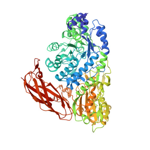
    8XRT, 8XRU, 8XRV, 8XRX

  • PubMed Abstract: 

    Glycoside hydrolases (GHs) are industrially important enzymes that hydrolyze glycosidic bonds in glycoconjugates. In this study, we found a GH3 β-glucosidase (CcBgl3B) from Cellulosimicrobium cellulans sp. 21 was able to selectively hydrolyze the β-1,6-glucosidic bond linked glucose of ginsenosides. X-ray crystallographic studies of the ligand complex ginsenoside-specific β-glucosidase provided a novel finding that support the catalytic mechanism of GH3. The substrate was clearly identified within the catalytic center of wild-type CcBgl3B, revealing that the C1 atom of the glucose was covalently bound to the Oδ1 group of the conserved catalytic nucleophile Asp264 as an enzyme-glycosyl intermediate. The glycosylated Asp264 could be identified by mass spectrometry. Through site-directed mutagenesis studies with Asp264, it was found that the covalent intermediate state formed by Asp264 and the substrate was critical for catalysis. In addition, Glu525 variants (E525A, E525Q and E525D) showed no or marginal activity against pNPβGlc; thus, this residue could supply a proton for the reaction. Overall, our study provides an insight into the catalytic mechanism of the GH3 enzyme CcBgl3B.


  • Organizational Affiliation
    • Engineering Research Center of Glycoconjugates Ministry of Education, Jilin Provincial Key Laboratory of Chemistry and Biology of Changbai Mountain Natural Drugs, School of Life Sciences, Northeast Normal University, Changchun 130024, China.

Macromolecule Content 

  • Total Structure Weight: 492.35 kDa 
  • Atom Count: 33,122 
  • Modeled Residue Count: 4,351 
  • Deposited Residue Count: 4,608 
  • Unique protein chains: 1

Macromolecules

Find similar proteins by:|  3D Structure
Entity ID: 1
MoleculeChains  Sequence LengthOrganismDetailsImage
GH3 enzyme CcBgl3B
A, B, C, D, E
A, B, C, D, E, F
768Cellulosimicrobium cellulansMutation(s): 0 
EC: 3.2.1.21
UniProt
Find proteins for A0AAT8XUL0 (Cellulosimicrobium cellulans)
Explore A0AAT8XUL0 
Go to UniProtKB:  A0AAT8XUL0
Entity Groups
Sequence Clusters30% Identity50% Identity70% Identity90% Identity95% Identity100% Identity
UniProt GroupA0AAT8XUL0
Sequence Annotations
Expand
Reference Sequence

Oligosaccharides

Help  
Entity ID: 2
MoleculeChains Length2D Diagram GlycosylationD Interactions
beta-D-glucopyranose-(1-6)-beta-D-glucopyranoseG,
H [auth J],
I [auth K]
2N/A
Glycosylation Resources
GlyTouCan: G09183JD
GlyCosmos: G09183JD

Small Molecules

Ligands 3 Unique
IDChains Name / Formula / InChI Key2D Diagram3D Interactions
BGC

Query on BGC


Download:Ideal Coordinates CCD File
K [auth B],
N [auth C]
beta-D-glucopyranose
C6 H12 O6
WQZGKKKJIJFFOK-VFUOTHLCSA-N
GLC

Query on GLC


Download:Ideal Coordinates CCD File
L [auth B]alpha-D-glucopyranose
C6 H12 O6
WQZGKKKJIJFFOK-DVKNGEFBSA-N
CA

Query on CA


Download:Ideal Coordinates CCD File
J [auth A]
M [auth B]
O [auth C]
P [auth D]
Q [auth E]
J [auth A],
M [auth B],
O [auth C],
P [auth D],
Q [auth E],
R [auth F]
CALCIUM ION
Ca
BHPQYMZQTOCNFJ-UHFFFAOYSA-N

Experimental Data & Validation

Experimental Data

  • Method: X-RAY DIFFRACTION
  • Resolution: 2.50 Å
  • R-Value Free:  0.230 (Depositor), 0.229 (DCC) 
  • R-Value Work:  0.197 (Depositor), 0.198 (DCC) 
  • R-Value Observed: 0.197 (Depositor) 
Space Group: C 1 2 1
Unit Cell:
Length ( Å )Angle ( ˚ )
a = 163.095α = 90
b = 183.009β = 100.59
c = 269.154γ = 90
Software Package:
Software NamePurpose
PHENIXrefinement

Structure Validation

View Full Validation Report


Ligand Structure Quality Assessment 


Entry History 

& Funding Information

Deposition Data

  • Released Date: 2024-04-10 
  • Deposition Author(s): Su, J.Y.

Funding OrganizationLocationGrant Number
National Natural Science Foundation of China (NSFC)China--

Revision History  (Full details and data files)

  • Version 1.0: 2024-04-10
    Type: Initial release