


The Crystal Structure of EHMT1 from Biortus.
Wang, F., Cheng, W., Yuan, Z., Lin, D., Bao, C.To be published.
Experimental Data Snapshot
Starting Model: experimental
View more details
Entity ID: 1 | |||||
|---|---|---|---|---|---|
| Molecule | Chains | Sequence Length | Organism | Details | Image |
| Histone-lysine N-methyltransferase EHMT1 | 285 | Homo sapiens | Mutation(s): 0 Gene Names: EHMT1, EUHMTASE1, GLP, KIAA1876, KMT1D EC: 2.1.1 (PDB Primary Data), 2.1.1.367 (PDB Primary Data) | 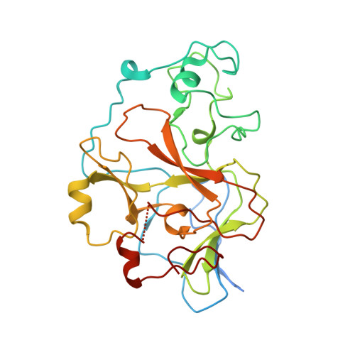 | |
UniProt & NIH Common Fund Data Resources | |||||
PHAROS: Q9H9B1 GTEx: ENSG00000181090 | |||||
Entity Groups | |||||
| Sequence Clusters | 30% Identity50% Identity70% Identity90% Identity95% Identity100% Identity | ||||
| UniProt Group | Q9H9B1 | ||||
Sequence AnnotationsExpand | |||||
Reference Sequence | |||||
| Ligands 3 Unique | |||||
|---|---|---|---|---|---|
| ID | Chains | Name / Formula / InChI Key | 2D Diagram | 3D Interactions | |
| SAH (Subject of Investigation/LOI) Download:Ideal Coordinates CCD File | E [auth A], J [auth B], Q [auth C], W [auth D] | S-ADENOSYL-L-HOMOCYSTEINE C14 H20 N6 O5 S ZJUKTBDSGOFHSH-WFMPWKQPSA-N |  | ||
| SO4 (Subject of Investigation/LOI) Download:Ideal Coordinates CCD File | BA [auth D], O [auth B], P [auth B], V [auth C] | SULFATE ION O4 S QAOWNCQODCNURD-UHFFFAOYSA-L |  | ||
| ZN (Subject of Investigation/LOI) Download:Ideal Coordinates CCD File | AA [auth D] F [auth A] G [auth A] H [auth A] I [auth A] | ZINC ION Zn PTFCDOFLOPIGGS-UHFFFAOYSA-N |  | ||
| Length ( Å ) | Angle ( ˚ ) |
|---|---|
| a = 85.113 | α = 90 |
| b = 94.638 | β = 112.337 |
| c = 88.731 | γ = 90 |
| Software Name | Purpose |
|---|---|
| REFMAC | refinement |
| XDS | data reduction |
| Aimless | data scaling |
| PHASER | phasing |


