


High Resolution Crystal Structure of the Pyruvate Kinase Tetramer in Complex with the Allosteric Activator Mitapivat/AG-348
Han, X., Sandalova, T., Zhang, C., Mardinoglu, A., Achour, A., Sun, R.(2024) Crystals (Basel)
Experimental Data Snapshot
Starting Model: experimental
View more details
(2024) Crystals (Basel)
Entity ID: 1 | |||||
|---|---|---|---|---|---|
| Molecule | Chains | Sequence Length | Organism | Details | Image |
| Isoform L-type of Pyruvate kinase PKLR | 543 | Homo sapiens | Mutation(s): 0 Gene Names: PKLR, PK1, PKL EC: 2.7.1.40 | 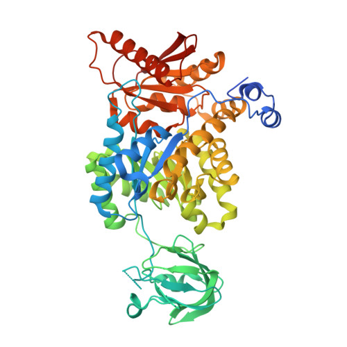 | |
UniProt & NIH Common Fund Data Resources | |||||
PHAROS: P30613 GTEx: ENSG00000143627 | |||||
Entity Groups | |||||
| Sequence Clusters | 30% Identity50% Identity70% Identity90% Identity95% Identity100% Identity | ||||
| UniProt Group | P30613 | ||||
Sequence AnnotationsExpand | |||||
Reference Sequence | |||||
| Ligands 3 Unique | |||||
|---|---|---|---|---|---|
| ID | Chains | Name / Formula / InChI Key | 2D Diagram | 3D Interactions | |
| WV2 Download:Ideal Coordinates CCD File | I [auth B], N [auth D] | mitapivat C24 H26 N4 O3 S XAYGBKHKBBXDAK-UHFFFAOYSA-N |  | ||
| FBP Download:Ideal Coordinates CCD File | F [auth A], H [auth B], K [auth C], M [auth D] | 1,6-di-O-phosphono-beta-D-fructofuranose C6 H14 O12 P2 RNBGYGVWRKECFJ-ARQDHWQXSA-N |  | ||
| MG Download:Ideal Coordinates CCD File | E [auth A], G [auth B], J [auth C], L [auth D] | MAGNESIUM ION Mg JLVVSXFLKOJNIY-UHFFFAOYSA-N |  | ||
| Length ( Å ) | Angle ( ˚ ) |
|---|---|
| a = 84.93 | α = 76.78 |
| b = 86.962 | β = 66.88 |
| c = 91.554 | γ = 80.22 |
| Software Name | Purpose |
|---|---|
| PHENIX | refinement |
| autoPROC | data processing |
| autoPROC | data reduction |
| Aimless | data scaling |
| PHASER | phasing |
| Coot | model building |
| Funding Organization | Location | Grant Number |
|---|---|---|
| Not funded | -- |


