8XET | pdb_00008xet

High-resolution structure of the siderophore periplasmic binding protein FtsB from Streptococcus pyogenes


Experimental Data Snapshot

  • Method: X-RAY DIFFRACTION
  • Resolution: 1.11 Å
  • R-Value Free: 
    0.142 (Depositor), 0.142 (DCC) 
  • R-Value Work: 
    0.131 (Depositor), 0.131 (DCC) 

Starting Model: experimental
View more details

wwPDB Validation 3D Report Full Report

Validation slider image for 8XET

This is version 1.2 of the entry. See complete history

Literature

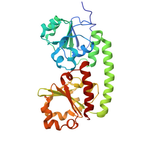
Structural basis for the ligand promiscuity of the hydroxamate siderophore binding protein FtsB from Streptococcus pyogenes.

Fernandez-Perez, J.Senoo, A.Caaveiro, J.M.M.Nakakido, M.de Vega, S.Nakagawa, I.Tsumoto, K.

(2024) Structure 32: 2410-2421.e3

  • DOI: https://doi.org/10.1016/j.str.2024.09.018
  • Primary Citation Related Structures: 
    8XET, 8XEU, 8XF4, 8XF7, 8XF8, 8XF9, 8XFA, 8XFI

  • PubMed Abstract: 

    Pathogenic bacteria must secure the uptake of nutritional metals such as iron for their growth, making their import systems attractive targets for the development of new antimicrobial modalities. In the pathogenic bacterium Streptococcus pyogenes, the iron uptake system FtsABCD transports iron encapsulated by siderophores of the hydroxamate class. However, the inability of S. pyogenes to produce these metabolites makes the biological and clinical relevance of this route unresolved. Herein, we demonstrated that the periplasmic binding protein FtsB recognizes not only the hydroxamate siderophore ferrichrome, as previously documented, but also ferrioxamine E (FOE), ferrioxamine B (FOB), and bisucaberin (BIS), each of them with high affinity (nM level). Up to seven aromatic residues in the binding pocket accommodate the variable backbones of the different siderophores through CH-π interactions, explaining ligand promiscuity. Collectively, our observations revealed how S. pyogenes exploits the diverse xenosiderophores produced by other microorganisms as iron sources to secure this precious nutrient.


  • Organizational Affiliation
    • Department of Bioengineering, School of Engineering, The University of Tokyo, 7-3-1 Hongo, Bunkyo-ku, Tokyo 113-8656, Japan.

Macromolecule Content 

  • Total Structure Weight: 32.9 kDa 
  • Atom Count: 2,753 
  • Modeled Residue Count: 279 
  • Deposited Residue Count: 283 
  • Unique protein chains: 1

Macromolecules

Find similar proteins by:|  3D Structure
Entity ID: 1
MoleculeChains  Sequence LengthOrganismDetailsImage
Iron-hydroxamate ABC transporter substrate-binding protein283Streptococcus pyogenes SSI-1Mutation(s): 0 
Gene Names: E0F66_02370
UniProt
Find proteins for A0A5S4TPK8 (Streptococcus pyogenes)
Explore A0A5S4TPK8 
Go to UniProtKB:  A0A5S4TPK8
Entity Groups
Sequence Clusters30% Identity50% Identity70% Identity90% Identity95% Identity100% Identity
UniProt GroupA0A5S4TPK8
Sequence Annotations
Expand
Reference Sequence

Small Molecules

Ligands 7 Unique
IDChains Name / Formula / InChI Key2D Diagram3D Interactions
P33

Query on P33


Download:Ideal Coordinates CCD File
B [auth A]3,6,9,12,15,18-HEXAOXAICOSANE-1,20-DIOL
C14 H30 O8
XPJRQAIZZQMSCM-UHFFFAOYSA-N
P6G

Query on P6G


Download:Ideal Coordinates CCD File
E [auth A]HEXAETHYLENE GLYCOL
C12 H26 O7
IIRDTKBZINWQAW-UHFFFAOYSA-N
PEG

Query on PEG


Download:Ideal Coordinates CCD File
C [auth A]DI(HYDROXYETHYL)ETHER
C4 H10 O3
MTHSVFCYNBDYFN-UHFFFAOYSA-N
ZN

Query on ZN


Download:Ideal Coordinates CCD File
F [auth A],
G [auth A],
H [auth A]
ZINC ION
Zn
PTFCDOFLOPIGGS-UHFFFAOYSA-N
EDO

Query on EDO


Download:Ideal Coordinates CCD File
D [auth A]1,2-ETHANEDIOL
C2 H6 O2
LYCAIKOWRPUZTN-UHFFFAOYSA-N
CL

Query on CL


Download:Ideal Coordinates CCD File
K [auth A]CHLORIDE ION
Cl
VEXZGXHMUGYJMC-UHFFFAOYSA-M
NA

Query on NA


Download:Ideal Coordinates CCD File
I [auth A],
J [auth A]
SODIUM ION
Na
FKNQFGJONOIPTF-UHFFFAOYSA-N

Experimental Data & Validation

Experimental Data

  • Method: X-RAY DIFFRACTION
  • Resolution: 1.11 Å
  • R-Value Free:  0.142 (Depositor), 0.142 (DCC) 
  • R-Value Work:  0.131 (Depositor), 0.131 (DCC) 
Space Group: P 65
Unit Cell:
Length ( Å )Angle ( ˚ )
a = 75.924α = 90
b = 75.924β = 90
c = 102.859γ = 120
Software Package:
Software NamePurpose
REFMACrefinement
XDSdata reduction
Aimlessdata scaling
PHASERphasing

Structure Validation

View Full Validation Report


Entry History 

& Funding Information

Deposition Data


Funding OrganizationLocationGrant Number
Japan Society for the Promotion of Science (JSPS)Japan20H02531

Revision History  (Full details and data files)

  • Version 1.0: 2024-10-09
    Type: Initial release
  • Version 1.1: 2024-10-30
    Changes: Database references
  • Version 1.2: 2024-12-18
    Changes: Database references