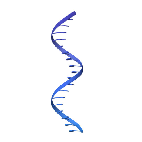
8XBY

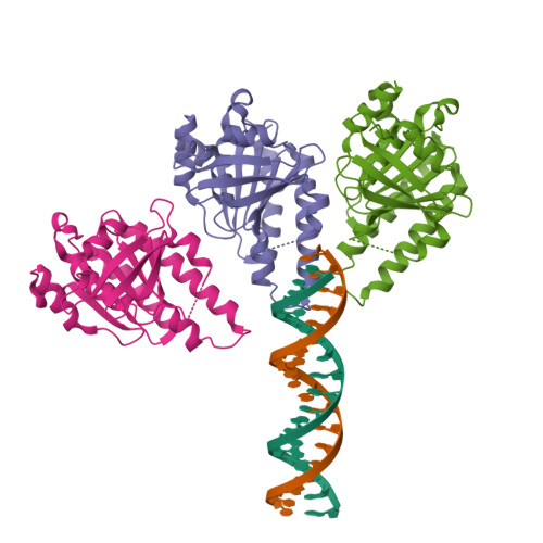
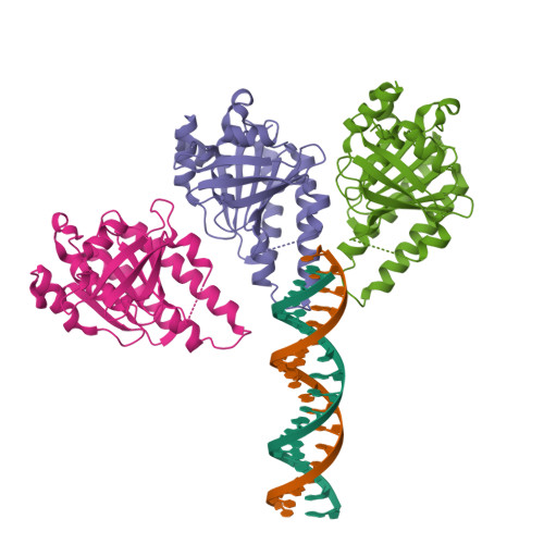
The cryo-EM structure of the RAD51 L1 and L2 loops bound to the linker DNA with the blunt end of the nucleosome


Experimental Data Snapshot

  • Method: ELECTRON MICROSCOPY
  • Resolution: 7.80 Å
  • Aggregation State: PARTICLE 
  • Reconstruction Method: SINGLE PARTICLE 

wwPDB Validation   3D Report Full Report


This is version 1.4 of the entry. See complete history


Literature

Cryo-EM structures of RAD51 assembled on nucleosomes containing a DSB site.

Shioi, T.Hatazawa, S.Oya, E.Hosoya, N.Kobayashi, W.Ogasawara, M.Kobayashi, T.Takizawa, Y.Kurumizaka, H.

(2024) Nature 628: 212-220

  • DOI: https://doi.org/10.1038/s41586-024-07196-4
  • Primary Citation of Related Structures:  
    8JND, 8JNE, 8JNF, 8XBT, 8XBU, 8XBV, 8XBW, 8XBX, 8XBY

  • PubMed Abstract: 

    RAD51 is the central eukaryotic recombinase required for meiotic recombination and mitotic repair of double-strand DNA breaks (DSBs) 1,2 . However, the mechanism by which RAD51 functions at DSB sites in chromatin has remained elusive. Here we report the cryo-electron microscopy structures of human RAD51-nucleosome complexes, in which RAD51 forms ring and filament conformations. In the ring forms, the N-terminal lobe domains (NLDs) of RAD51 protomers are aligned on the outside of the RAD51 ring, and directly bind to the nucleosomal DNA. The nucleosomal linker DNA that contains the DSB site is recognized by the L1 and L2 loops-active centres that face the central hole of the RAD51 ring. In the filament form, the nucleosomal DNA is peeled by the RAD51 filament extension, and the NLDs of RAD51 protomers proximal to the nucleosome bind to the remaining nucleosomal DNA and histones. Mutations that affect nucleosome-binding residues of the RAD51 NLD decrease nucleosome binding, but barely affect DNA binding in vitro. Consistently, yeast Rad51 mutants with the corresponding mutations are substantially defective in DNA repair in vivo. These results reveal an unexpected function of the RAD51 NLD, and explain the mechanism by which RAD51 associates with nucleosomes, recognizes DSBs and forms the active filament in chromatin.


  • Organizational Affiliation

    Laboratory of Chromatin Structure and Function, Institute for Quantitative Biosciences, The University of Tokyo, Tokyo, Japan.


Macromolecules

Find similar proteins by:  (by identity cutoff)  |  3D Structure
Entity ID: 3
MoleculeChains Sequence LengthOrganismDetailsImage
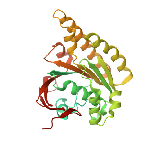
DNA repair protein RAD51 homolog 1C [auth K],
D [auth L],
E [auth M]
342Homo sapiensMutation(s): 0 
Gene Names: RAD51RAD51ARECA
UniProt & NIH Common Fund Data Resources
Find proteins for Q06609 (Homo sapiens)
Explore Q06609 
Go to UniProtKB:  Q06609
PHAROS:  Q06609
GTEx:  ENSG00000051180 
Entity Groups  
Sequence Clusters30% Identity50% Identity70% Identity90% Identity95% Identity100% Identity
UniProt GroupQ06609
Sequence Annotations
Expand
  • Reference Sequence
Find similar nucleic acids by:  (by identity cutoff)  |  3D Structure
Entity ID: 1
MoleculeChains LengthOrganismImage
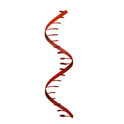
DNA (5'-D(P*AP*AP*CP*GP*AP*AP*AP*AP*CP*GP*GP*CP*CP*AP*CP*CP*AP*CP*G)-3')A [auth I]158synthetic construct
Sequence Annotations
Expand
  • Reference Sequence
Find similar nucleic acids by:  (by identity cutoff)  |  3D Structure
Entity ID: 2
MoleculeChains LengthOrganismImage
DNA (5'-D(P*CP*GP*TP*GP*GP*TP*GP*GP*CP*CP*GP*TP*TP*TP*TP*CP*GP*TP*T)-3')B [auth J]158synthetic construct
Sequence Annotations
Expand
  • Reference Sequence
Experimental Data & Validation

Experimental Data

  • Method: ELECTRON MICROSCOPY
  • Resolution: 7.80 Å
  • Aggregation State: PARTICLE 
  • Reconstruction Method: SINGLE PARTICLE 

Structure Validation

View Full Validation Report


Entry History & Funding Information

Deposition Data


Funding OrganizationLocationGrant Number
Japan Society for the Promotion of Science (JSPS)JapanJP22K06098
Japan Society for the Promotion of Science (JSPS)JapanJP23H05475
Japan Agency for Medical Research and Development (AMED)JapanJP23ama121009
Japan Science and TechnologyJapanJPMJER1901
Japan Society for the Promotion of Science (JSPS)JapanJP23K14134

Revision History  (Full details and data files)

  • Version 1.0: 2024-03-27
    Type: Initial release
  • Version 1.1: 2024-04-03
    Changes: Database references
  • Version 1.2: 2024-04-17
    Changes: Database references
  • Version 1.3: 2024-04-24
    Changes: Database references
  • Version 1.4: 2024-05-08
    Changes: Database references