


Decarboxylative Vanillate 1-Hydroxylase from Phanerochaete chrysosporium
Mori, R., Suzuki, H., Ishida, T., Igarashi, K., Kato, M., Shimizu, M.To be published.
Experimental Data Snapshot
Starting Model: in silico
View more details
Entity ID: 1 | |||||
|---|---|---|---|---|---|
| Molecule | Chains | Sequence Length | Organism | Details | Image |
| Decarboxylative Vanillate 1-Hydroxylase | 496 | Phanerochaete chrysosporium RP-78 | Mutation(s): 0 Gene Names: fgenesh1_kg.3_#_10694_#_transcript/44524 EC: 1.14.13.1 | 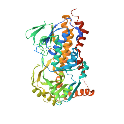 | |
| Ligands 2 Unique | |||||
|---|---|---|---|---|---|
| ID | Chains | Name / Formula / InChI Key | 2D Diagram | 3D Interactions | |
| FAD (Subject of Investigation/LOI) Download:Ideal Coordinates CCD File | B [auth A] | FLAVIN-ADENINE DINUCLEOTIDE C27 H33 N9 O15 P2 VWWQXMAJTJZDQX-UYBVJOGSSA-N |  | ||
| ACT Download:Ideal Coordinates CCD File | C [auth A] | ACETATE ION C2 H3 O2 QTBSBXVTEAMEQO-UHFFFAOYSA-M |  | ||
| Length ( Å ) | Angle ( ˚ ) |
|---|---|
| a = 45.632 | α = 90 |
| b = 97.13 | β = 90 |
| c = 106.596 | γ = 90 |
| Software Name | Purpose |
|---|---|
| REFMAC | refinement |
| XDS | data reduction |
| Aimless | data scaling |
| MOLREP | phasing |
| Funding Organization | Location | Grant Number |
|---|---|---|
| Japan Society for the Promotion of Science (JSPS) | Japan | 20K05815 |


