8X22 | pdb_00008x22

HIV-1 reverse transcriptase mutant Q151M/Y115F/F116Y/L74V:DNA:dGTP ternary complex


Experimental Data Snapshot

  • Method: X-RAY DIFFRACTION
  • Resolution: 2.31 Å
  • R-Value Free: 
    0.219 (Depositor), 0.222 (DCC) 
  • R-Value Work: 
    0.182 (Depositor), 0.185 (DCC) 
  • R-Value Observed: 
    0.184 (Depositor) 

Starting Model: experimental
View more details

wwPDB Validation 3D Report Full Report

Validation slider image for 8X22

Ligand Structure Quality Assessment 


This is version 1.0 of the entry. See complete history

Literature

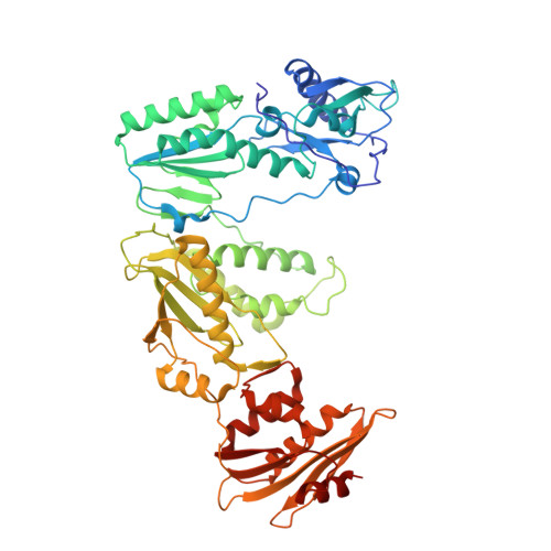
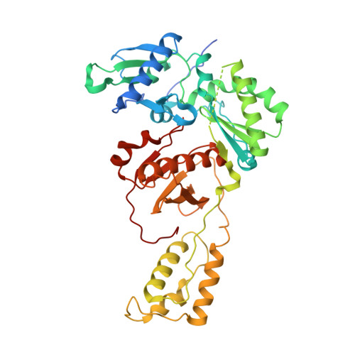
Deviated binding of anti-HBV nucleoside analog E-CFCP-TP to the reverse transcriptase active site attenuates the effect of drug-resistant mutations.

Yasutake, Y.Hattori, S.I.Kumamoto, H.Tamura, N.Maeda, K.Mitsuya, H.

(2024) Sci Rep 14: 15742-15742

  • DOI: https://doi.org/10.1038/s41598-024-66505-z
  • Primary Citation Related Structures: 
    8X1Z, 8X20, 8X21, 8X22

  • PubMed Abstract: 

    While certain human hepatitis B virus-targeting nucleoside analogs (NAs) serve as crucial anti-HBV drugs, HBV yet remains to be a major global health threat. E-CFCP is a 4'-modified and fluoromethylenated NA that exhibits potent antiviral activity against both wild-type and drug-resistant HBVs but less potent against human immunodeficiency virus type-1 (HIV-1). Here, we show that HIV-1 with HBV-associated amino acid substitutions introduced into the RT's dNTP-binding site (N-site) is highly susceptible to E-CFCP. We determined the X-ray structures of HBV-associated HIV-1 RT mutants complexed with DNA:E-CFCP-triphosphate (E-CFCP-TP). The structures revealed that exocyclic fluoromethylene pushes the Met184 sidechain backward, and the resultant enlarged hydrophobic pocket accommodates both the fluoromethylene and 4'-cyano moiety of E-CFCP. Structural comparison with the DNA:dGTP/entecavir-triphosphate complex also indicated that the cyclopentene moiety of the bound E-CFCP-TP is slightly skewed and deviated. This positioning partly corresponds to that of the bound dNTP observed in the HIV-1 RT mutant with drug-resistant mutations F160M/M184V, resulting in the attenuation of the structural effects of F160M/M184V substitutions. These results expand our knowledge of the interactions between NAs and the RT N-site and should help further design antiviral NAs against both HIV-1 and HBV.


  • Organizational Affiliation
    • Bioproduction Research Institute, National Institute of Advanced Industrial Science and Technology (AIST), Sapporo, 062-8517, Japan. y-yasutake@aist.go.jp.

Macromolecule Content 

  • Total Structure Weight: 256.83 kDa 
  • Atom Count: 17,744 
  • Modeled Residue Count: 1,991 
  • Deposited Residue Count: 2,078 
  • Unique protein chains: 2
  • Unique nucleic acid chains: 1

Macromolecules


Find similar proteins by:|  3D Structure
Entity ID: 1
MoleculeChains  Sequence LengthOrganismDetailsImage
Pol protein (Fragment)A,
D [auth C]
557Human immunodeficiency virus 1Mutation(s): 6 
Gene Names: pol
UniProt
Find proteins for P12497 (Human immunodeficiency virus type 1 group M subtype B (isolate NY5))
Explore P12497 
Go to UniProtKB:  P12497
Entity Groups
Sequence Clusters30% Identity50% Identity70% Identity90% Identity95% Identity100% Identity
UniProt GroupP12497
Sequence Annotations
Expand
Reference Sequence
Find similar proteins by:|  3D Structure
Entity ID: 2
MoleculeChains  Sequence LengthOrganismDetailsImage
HIV-1 RT p51 subunitB,
E [auth D]
444Human immunodeficiency virus 1Mutation(s): 2 
UniProt
Find proteins for P12497 (Human immunodeficiency virus type 1 group M subtype B (isolate NY5))
Explore P12497 
Go to UniProtKB:  P12497
Entity Groups
Sequence Clusters30% Identity50% Identity70% Identity90% Identity95% Identity100% Identity
UniProt GroupP12497
Sequence Annotations
Expand
Reference Sequence
Find similar nucleic acids by:  (by identity cutoff) 
Entity ID: 3
MoleculeChains LengthOrganismImage
DNA/RNA (38-MER)C [auth E],
F
38synthetic construct
Sequence Annotations
Expand
Reference Sequence

Small Molecules

Ligands 3 Unique
IDChains Name / Formula / InChI Key2D Diagram3D Interactions
DGT
(Subject of Investigation/LOI)

Query on DGT


Download:Ideal Coordinates CCD File
G [auth A],
L [auth C]
2'-DEOXYGUANOSINE-5'-TRIPHOSPHATE
C10 H16 N5 O13 P3
HAAZLUGHYHWQIW-KVQBGUIXSA-N
GOL

Query on GOL


Download:Ideal Coordinates CCD File
I [auth A]
J [auth A]
K [auth B]
N [auth C]
O [auth D]
I [auth A],
J [auth A],
K [auth B],
N [auth C],
O [auth D],
P [auth D]
GLYCEROL
C3 H8 O3
PEDCQBHIVMGVHV-UHFFFAOYSA-N
MG

Query on MG


Download:Ideal Coordinates CCD File
H [auth A],
M [auth C]
MAGNESIUM ION
Mg
JLVVSXFLKOJNIY-UHFFFAOYSA-N

Experimental Data & Validation

Experimental Data

  • Method: X-RAY DIFFRACTION
  • Resolution: 2.31 Å
  • R-Value Free:  0.219 (Depositor), 0.222 (DCC) 
  • R-Value Work:  0.182 (Depositor), 0.185 (DCC) 
  • R-Value Observed: 0.184 (Depositor) 
Space Group: H 3
Unit Cell:
Length ( Å )Angle ( ˚ )
a = 285.227α = 90
b = 285.227β = 90
c = 96.075γ = 120
Software Package:
Software NamePurpose
PHENIXrefinement
XDSdata reduction
Aimlessdata scaling
MOLREPphasing

Structure Validation

View Full Validation Report


Ligand Structure Quality Assessment 


Entry History 

& Funding Information

Deposition Data


Funding OrganizationLocationGrant Number
Japan Agency for Medical Research and Development (AMED)JapanJP21fk0310113

Revision History  (Full details and data files)

  • Version 1.0: 2024-07-17
    Type: Initial release