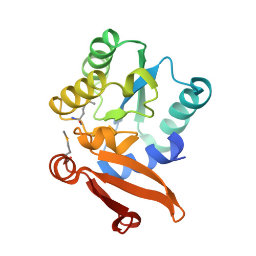
8WXN

Crystal structure of substrate-binding protein from Rhodothermus marinus (Dose II)


Experimental Data Snapshot

  • Method: X-RAY DIFFRACTION
  • Resolution: 1.58 Å
  • R-Value Free: 0.259 
  • R-Value Work: 0.222 
  • R-Value Observed: 0.224 

Starting Model: experimental
View more details

wwPDB Validation   3D Report Full Report


This is version 1.3 of the entry. See complete history


Literature

Data of radiation damage on selenomethionine-substituted single-domain substrate-binding protein.

Nam, K.H.

(2024) Data Brief 53: 110114-110114

  • DOI: https://doi.org/10.1016/j.dib.2024.110114
  • Primary Citation of Related Structures:  
    8WXM, 8WXN, 8WXO, 8WXP

  • PubMed Abstract: 

    Radiation damage is an inherent issue in X-ray crystallography. It not only damages macromolecular crystals, which lowers the quality of the diffraction intensity, but results in inaccurate structural information. Among the various types of radiation damage, little is known regarding the damage to selenomethionine, an amino acid contained in some proteins. Recently, radiation damage to the selenomethionine-substituted single-domain substrate-binding domain from Rhodothermus marinus (SeMet-RmSBP) was investigated. Global and specific radiation damage from four datasets collected by repeatedly exposing a single RmSBP-SeMet crystal to X-rays were analyzed. The results indicated that the B-factor value of the selenium atom in selenomethionine was significantly increased compared with other atoms. To date, no images of radiation damage have been reported for selenomethionine-substituted proteins. Therefore, these data may be used to study radiation damage in macromolecular crystallography. This study provides insight into radiation damage associated with selenomethionine.


  • Organizational Affiliation

    College of General Education, Kookmin University, Seoul 02707, South Korea.


Macromolecules
Find similar proteins by:  (by identity cutoff)  |  3D Structure
Entity ID: 1
MoleculeChains Sequence LengthOrganismDetailsImage
ABC-type uncharacterized transport system periplasmic component-like protein
A, B
164Rhodothermus marinus DSM 4252Mutation(s): 0 
Gene Names: Rmar_2176
UniProt
Find proteins for D0MDR1 (Rhodothermus marinus (strain ATCC 43812 / DSM 4252 / R-10))
Explore D0MDR1 
Go to UniProtKB:  D0MDR1
Entity Groups  
Sequence Clusters30% Identity50% Identity70% Identity90% Identity95% Identity100% Identity
UniProt GroupD0MDR1
Sequence Annotations
Expand
  • Reference Sequence
Small Molecules
Modified Residues  1 Unique
IDChains TypeFormula2D DiagramParent
MSE
Query on MSE
A, B
L-PEPTIDE LINKINGC5 H11 N O2 SeMET
Experimental Data & Validation

Experimental Data

  • Method: X-RAY DIFFRACTION
  • Resolution: 1.58 Å
  • R-Value Free: 0.259 
  • R-Value Work: 0.222 
  • R-Value Observed: 0.224 
  • Space Group: C 1 2 1
Unit Cell:
Length ( Å )Angle ( ˚ )
a = 63.552α = 90
b = 64.164β = 96.07
c = 69.863γ = 90
Software Package:
Software NamePurpose
PHENIXrefinement
HKL-2000data reduction
HKL-2000data scaling
MOLREPphasing

Structure Validation

View Full Validation Report


Entry History & Funding Information

Deposition Data

  • Released Date: 2023-11-22 
  • Deposition Author(s): Nam, K.H.

Funding OrganizationLocationGrant Number
National Research Foundation (NRF, Korea)Korea, Republic OfNRF-2017M3A9F6029736
National Research Foundation (NRF, Korea)Korea, Republic OfNRF-2021R1I1A1A01050838
National Research Foundation (NRF, Korea)Korea, Republic OfNRF-2020M3H1A1075314

Revision History  (Full details and data files)

  • Version 1.0: 2023-11-22
    Type: Initial release
  • Version 1.1: 2024-04-17
    Changes: Database references
  • Version 1.2: 2024-05-22
    Changes: Database references
  • Version 1.3: 2024-10-23
    Changes: Structure summary