8WWY | pdb_00008wwy

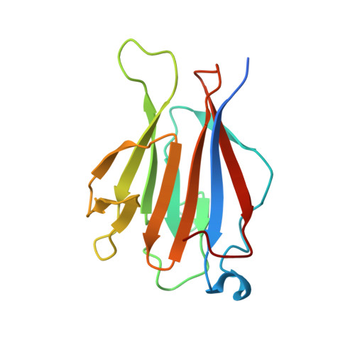
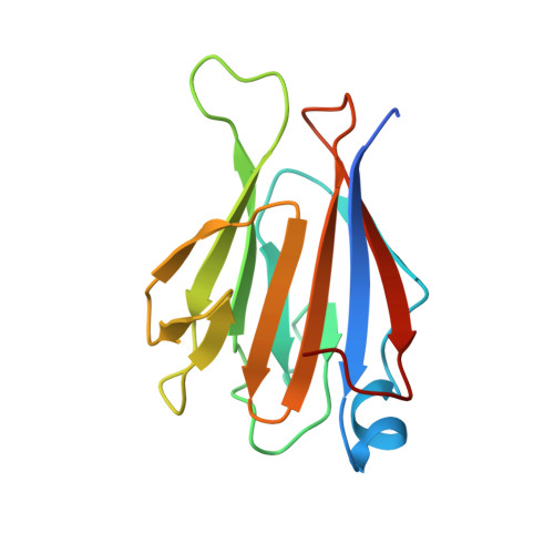
Crystal structure of mouse TIFA/TIFAB heterodimer


Experimental Data Snapshot

  • Method: X-RAY DIFFRACTION
  • Resolution: 1.79 Å
  • R-Value Free: 
    0.225 (Depositor), 0.226 (DCC) 
  • R-Value Work: 
    0.181 (Depositor), 0.183 (DCC) 
  • R-Value Observed: 
    0.183 (Depositor) 

Starting Model: experimental
View more details

wwPDB Validation 3D Report Full Report

Validation slider image for 8WWY

This is version 1.0 of the entry. See complete history. 

Literature

TIFAB regulates the TIFA-TRAF6 signaling pathway involved in innate immunity by forming a heterodimer complex with TIFA.

Nakamura, T., Ohyama, C., Sakamoto, M., Toma, T., Tateishi, H., Matsuo, M., Chirifu, M., Ikemizu, S., Morioka, H., Fujita, M., Inoue, J.I., Yamagata, Y.

(2024) Proc Natl Acad Sci U S A 121: e2318794121-e2318794121

  • DOI: https://doi.org/10.1073/pnas.2318794121
  • Primary Citation Related Structures: 
    8WWY

  • PubMed Abstract: 

    Nuclear factor κB (NF-κB) is activated by various inflammatory and infectious molecules and is involved in immune responses. It has been elucidated that ADP-β-D-manno-heptose (ADP-Hep), a metabolite in gram-negative bacteria, activates NF-κB through alpha-kinase 1 (ALPK1)-TIFA-TRAF6 signaling. ADP-Hep stimulates the kinase activity of ALPK1 for TIFA phosphorylation. Complex formation between phosphorylation-dependent TIFA oligomer and TRAF6 promotes the polyubiquitination of TRAF6 for NF-κB activation. TIFAB, a TIFA homolog lacking a phosphorylation site and a TRAF6 binding motif, is a negative regulator of TIFA-TRAF6 signaling and is implicated in myeloid diseases. TIFAB is indicated to regulate TIFA-TRAF6 signaling through interactions with TIFA and TRAF6; however, little is known about its biological function. We demonstrated that TIFAB forms a complex not with the TIFA dimer, an intrinsic form of TIFA involved in NF-κB activation, but with monomeric TIFA. The structural analysis of the TIFA/TIFAB complex and the biochemical and cell-based analyses showed that TIFAB forms a stable heterodimer with TIFA, inhibits TIFA dimer formation, and suppresses TIFA-TRAF6 signaling. The resultant TIFA/TIFAB complex is a "pseudo-TIFA dimer" lacking the phosphorylation site and TRAF6 binding motif in TIFAB and cannot form the orderly structure as proposed for the phosphorylated TIFA oligomer involved in NF-κB activation. This study elucidated the molecular and structural basis for the regulation of TIFA-TRAF6 signaling by TIFAB.


  • Organizational Affiliation: 
    • Graduate School of Pharmaceutical Sciences, Kumamoto University, Kumamoto 862-0973, Japan.

Macromolecule Content 

  • Total Structure Weight: 67.96 kDa 
  • Atom Count: 5,101 
  • Modeled Residue Count: 548 
  • Deposited Residue Count: 588 
  • Unique protein chains: 2

Macromolecules

Find similar proteins by:|  3D Structure
Entity ID: 1
MoleculeChains  Sequence LengthOrganismDetailsImage
TRAF-interacting protein with FHA domain-containing protein A
A, C
150Mus musculusMutation(s): 1 
Gene Names: Tifa
UniProt
Find proteins for Q793I8 (Mus musculus)
Explore Q793I8 
Go to UniProtKB:  Q793I8
Entity Groups
Sequence Clusters30% Identity50% Identity70% Identity90% Identity95% Identity100% Identity
UniProt GroupQ793I8
Sequence Annotations
Expand
Reference Sequence
Find similar proteins by:|  3D Structure
Entity ID: 2
MoleculeChains  Sequence LengthOrganismDetailsImage
TRAF-interacting protein with FHA domain-containing protein B
B, D
144Mus musculusMutation(s): 0 
Gene Names: Tifab
UniProt & NIH Common Fund Data Resources
Find proteins for Q8JZM6 (Mus musculus)
Explore Q8JZM6 
Go to UniProtKB:  Q8JZM6
IMPC:  MGI:2385852
Entity Groups
Sequence Clusters30% Identity50% Identity70% Identity90% Identity95% Identity100% Identity
UniProt GroupQ8JZM6
Sequence Annotations
Expand
Reference Sequence

Experimental Data & Validation

Experimental Data

  • Method: X-RAY DIFFRACTION
  • Resolution: 1.79 Å
  • R-Value Free:  0.225 (Depositor), 0.226 (DCC) 
  • R-Value Work:  0.181 (Depositor), 0.183 (DCC) 
  • R-Value Observed: 0.183 (Depositor) 
Space Group: P 61 2 2
Unit Cell:
Length ( Å )Angle ( ˚ )
a = 88.758α = 90
b = 88.758β = 90
c = 297.367γ = 120
Software Package:
Software NamePurpose
PHENIXrefinement
XDSdata reduction
XDSdata scaling
MOLREPphasing

Structure Validation

View Full Validation Report


Entry History 

& Funding Information

Deposition Data

  • Released Date: 2024-03-13 
  • Deposition Author(s): Nakamura, T.

Funding OrganizationLocationGrant Number
Japan Society for the Promotion of Science (JSPS)Japan--

Revision History  (Full details and data files)

  • Version 1.0: 2024-03-13
    Type: Initial release