Thermotolerance Mechanism of Fungal GH6 Cellobiohydrolase. Part II. Structural Analysis of Thermotolerant Mutant from the Basidiomycete Phanerochaete chrysosporium.
Yamaguchi, S., Sunagawa, N., Samejima, M., Igarashi, K.(2024) J Appl Glycosci (1999) 71: 63-72
- PubMed: 38863950 Search on PubMedSearch on PubMed Central
- DOI: https://doi.org/10.5458/jag.jag.JAG-2023_0018
- Primary Citation Related Structures:
8WUP, 8WW5, 8WWT, 8WX6 - PubMed Abstract:
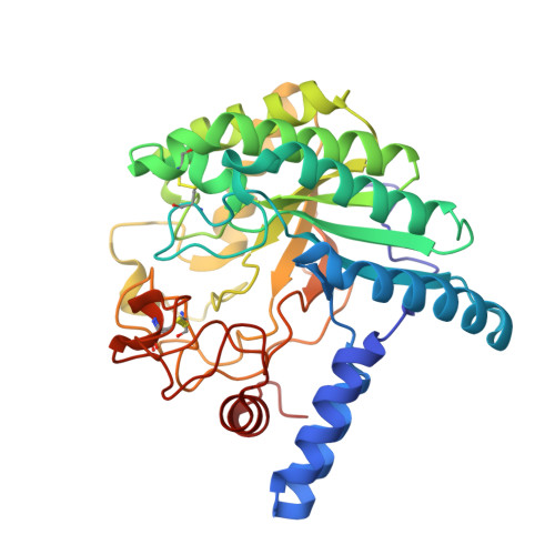
Glycoside hydrolase family 6 cellobiohydrolase (GH6 CBH) is a group of cellulases capable of hydrolyzing crystalline cellulose. However, the synergistic reaction of GH6 CBH with other cellulases is hindered by its relatively low thermotolerance. We previously obtained a thermotolerant double mutant, C240S/C393S, of GH6 CBH from the basidiomycete Phanerochaete chrysosporium ( Pc Cel6A) by replacing the two free cysteine (Cys) residues, C240 and C393, with serine (Yamaguchi et al ., J Appl Glycosci. 2020; 67;79-86). In the accompanying paper (Part I; Yamaguchi et al ., J Appl Glycosci. 2024; 71: 55-62), we measured the temperature dependence of the activity and folding of C240S/C393S and its single mutants, C240S and C393S, and found that replacement of C393 was the major contributor to the increased thermotolerance of C240S/C393S. Here, in order to investigate the mechanism involved, we crystallized the wild-type and the mutant enzymes and compared their X-ray crystal structures. The overall structures of the wild-type and the three mutant enzymes were similar. However, C240S/C393S had the lowest relative B -factor at both the N-terminal loop (residues 172-177) and the C-terminal loop (residues 390-425). This result suggests that reduced structural fluctuation of the substrate-enclosing loops, possibly due to stronger hydrogen bonding involving C393, could account for the increased thermotolerance of C240S/C393S.
- 1 Department of Biomaterial Sciences, Graduate School of Agricultural and Life Sciences, The University of Tokyo.
Organizational Affiliation: