8WP8 | pdb_00008wp8

Cryo-EM structure of SARS-CoV-2 Omicron BA.2.86 RBD in complex with human ACE2


Experimental Data Snapshot

  • Method: ELECTRON MICROSCOPY
  • Resolution: 2.89 Å
  • Aggregation State: PARTICLE 
  • Reconstruction Method: SINGLE PARTICLE 

wwPDB Validation 3D Report Full Report

Validation slider image for 8WP8

This is version 1.2 of the entry. See complete history

Literature

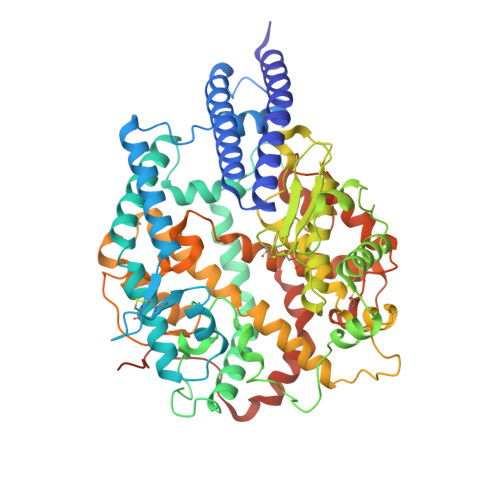
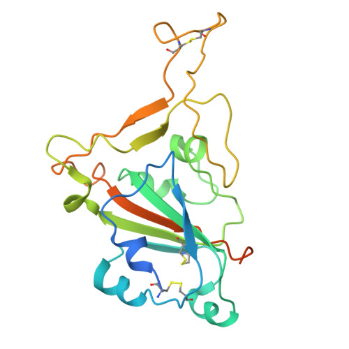
Spike structures, receptor binding, and immune escape of recently circulating SARS-CoV-2 Omicron BA.2.86, JN.1, EG.5, EG.5.1, and HV.1 sub-variants.

Li, L.Shi, K.Gu, Y.Xu, Z.Shu, C.Li, D.Sun, J.Cong, M.Li, X.Zhao, X.Yu, G.Hu, S.Tan, H.Qi, J.Ma, X.Liu, K.Gao, G.F.

(2024) Structure 32: 1055-1067.e6

  • DOI: https://doi.org/10.1016/j.str.2024.06.012
  • Primary Citation Related Structures: 
    8WP8, 8XLV, 8XM5, 8XMG, 8XMT, 8XN2, 8XN3, 8XN5, 8XNF, 8XNK, 8Y16, 8Y18, 8Y5J, 8Y6A

  • PubMed Abstract: 

    The recently emerged BA.2.86, JN.1, EG.5, EG.5.1, and HV.1 variants have a growth advantage. In this study, we explore the structural bases of receptor binding and immune evasion for the Omicron BA.2.86, JN.1, EG.5, EG.5.1, and HV.1 sub-variants. Our findings reveal that BA.2.86 exhibits strong receptor binding, whereas its JN.1 sub-lineage displays a decreased binding affinity to human ACE2 (hACE2). Through complex structure analyses, we observed that the reversion of R493Q in BA.2.86 receptor binding domain (RBD) plays a facilitating role in receptor binding, while the L455S substitution in JN.1 RBD restores optimal affinity. Furthermore, the structure of monoclonal antibody (mAb) S309 complexed with BA.2.86 RBD highlights the importance of the K356T mutation, which brings a new N-glycosylation motif, altering the binding pattern of mAbs belonging to RBD-5 represented by S309. These findings emphasize the importance of closely monitoring BA.2.86 and its sub-lineages to prevent another wave of SARS-CoV-2 infections.


  • Organizational Affiliation
    • CAS Key Laboratory of Pathogen Microbiology and Immunology, Institute of Microbiology, Chinese Academy of Sciences (CAS), Beijing, China; Beijing Life Science Academy, Beijing, China.

Macromolecule Content 

  • Total Structure Weight: 99.28 kDa 
  • Atom Count: 6,539 
  • Modeled Residue Count: 780 
  • Deposited Residue Count: 834 
  • Unique protein chains: 2

Macromolecules

Find similar proteins by:|  3D Structure
Entity ID: 1
MoleculeChains  Sequence LengthOrganismDetailsImage
Processed angiotensin-converting enzyme 2606Homo sapiensMutation(s): 0 
Gene Names: ACE2UNQ868/PRO1885
EC: 3.4.17 (UniProt), 3.4.17.23 (UniProt)
UniProt & NIH Common Fund Data Resources
Find proteins for Q9BYF1 (Homo sapiens)
Explore Q9BYF1 
Go to UniProtKB:  Q9BYF1
PHAROS:  Q9BYF1
GTEx:  ENSG00000130234 
Entity Groups
Sequence Clusters30% Identity50% Identity70% Identity90% Identity95% Identity100% Identity
UniProt GroupQ9BYF1
Glycosylation
Glycosylation Sites: 6Go to GlyGen: Q9BYF1-1
Sequence Annotations
Expand
Reference Sequence
Find similar proteins by:|  3D Structure
Entity ID: 2
MoleculeChains  Sequence LengthOrganismDetailsImage
Spike protein S1228Severe acute respiratory syndrome coronavirus 2Mutation(s): 18 
Gene Names: S2
UniProt
Find proteins for P0DTC2 (Severe acute respiratory syndrome coronavirus 2)
Explore P0DTC2 
Go to UniProtKB:  P0DTC2
Entity Groups
Sequence Clusters30% Identity50% Identity70% Identity90% Identity95% Identity100% Identity
UniProt GroupP0DTC2
Glycosylation
Glycosylation Sites: 2Go to GlyGen: P0DTC2-1
Sequence Annotations
Expand
Reference Sequence

Oligosaccharides

Help  
Entity ID: 3
MoleculeChains Length2D Diagram GlycosylationD Interactions
beta-D-mannopyranose-(1-4)-2-acetamido-2-deoxy-beta-D-glucopyranose-(1-4)-2-acetamido-2-deoxy-beta-D-glucopyranose
C, E
3N-Glycosylation
Glycosylation Resources
GlyTouCan: G15407YE
GlyCosmos: G15407YE
GlyGen: G15407YE
Entity ID: 4
MoleculeChains Length2D Diagram GlycosylationD Interactions
2-acetamido-2-deoxy-beta-D-glucopyranose-(1-4)-2-acetamido-2-deoxy-beta-D-glucopyranose
D
2N-Glycosylation
Glycosylation Resources
GlyTouCan: G42666HT
GlyCosmos: G42666HT
GlyGen: G42666HT

Experimental Data & Validation

Experimental Data

  • Method: ELECTRON MICROSCOPY
  • Resolution: 2.89 Å
  • Aggregation State: PARTICLE 
  • Reconstruction Method: SINGLE PARTICLE 
EM Software:
TaskSoftware PackageVersion
MODEL REFINEMENTPHENIX1.20.1_4487:

Structure Validation

View Full Validation Report


Entry History 

& Funding Information

Deposition Data


Funding OrganizationLocationGrant Number
Chinese Academy of SciencesChinaXDB29010202

Revision History  (Full details and data files)

  • Version 1.0: 2024-07-03
    Type: Initial release
  • Version 1.1: 2024-11-06
    Changes: Data collection, Structure summary
  • Version 1.2: 2025-07-16
    Changes: Data collection, Database references