


Structural insights into glycoside hydrolase family 1 beta-glucosidase: Selective oligosaccharide hydrolysis, synthesis, and product profiling.
Lin, C.-C., Uno, H., Yamada, C., Terada, T., Lu, T.-J., Fushinobu, S.(2026) J Biological Chem
Experimental Data Snapshot
(2026) J Biological Chem
Entity ID: 1 | |||||
|---|---|---|---|---|---|
| Molecule | Chains | Sequence Length | Organism | Details | Image |
| beta-glucosidase Td2F2 | 465 | metagenomes | Mutation(s): 0 EC: 3.2.1.21 | 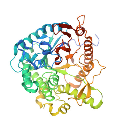 | |
| Ligands 3 Unique | |||||
|---|---|---|---|---|---|
| ID | Chains | Name / Formula / InChI Key | 2D Diagram | 3D Interactions | |
| NHE Download:Ideal Coordinates CCD File | D [auth A] | 2-[N-CYCLOHEXYLAMINO]ETHANE SULFONIC ACID C8 H17 N O3 S MKWKNSIESPFAQN-UHFFFAOYSA-N |  | ||
| BGC (Subject of Investigation/LOI) Download:Ideal Coordinates CCD File | C [auth A] | beta-D-glucopyranose C6 H12 O6 WQZGKKKJIJFFOK-VFUOTHLCSA-N |  | ||
| MLA Download:Ideal Coordinates CCD File | B [auth A] | MALONIC ACID C3 H4 O4 OFOBLEOULBTSOW-UHFFFAOYSA-N |  | ||
| Length ( Å ) | Angle ( ˚ ) |
|---|---|
| a = 67.608 | α = 90 |
| b = 69.24 | β = 90 |
| c = 96.683 | γ = 90 |
| Software Name | Purpose |
|---|---|
| REFMAC | refinement |
| Aimless | data scaling |
| XDS | data reduction |
| PHASER | phasing |
| Funding Organization | Location | Grant Number |
|---|---|---|
| Not funded | -- |


