


The structure of D-mandelate dehydrogenase with L103G and T143G mutations
Liu, F., Rao, Z.M.To be published.
Experimental Data Snapshot
Starting Model: experimental
View more details
wwPDB Validation 3D Report Full Report

Entity ID: 1 | |||||
|---|---|---|---|---|---|
| Molecule | Chains | Sequence Length | Organism | Details | Image |
| 2-dehydropantoate 2-reductase | 312 | Levilactobacillus brevis | Mutation(s): 2 Gene Names: CNR30_12720, UCCLB556_2064, LbMDH EC: 1.1.1.169 | 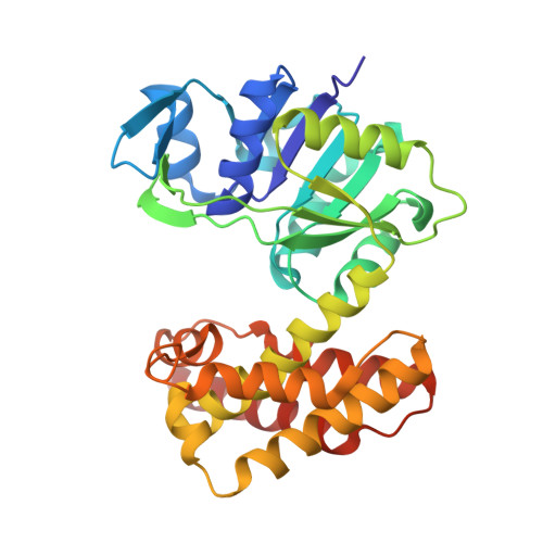 | |
| Length ( Å ) | Angle ( ˚ ) |
|---|---|
| a = 82.62 | α = 90 |
| b = 131.99 | β = 90 |
| c = 58.64 | γ = 90 |
| Software Name | Purpose |
|---|---|
| REFMAC | refinement |
| XDS | data reduction |
| HKL-3000 | data scaling |
| HKL-3000 | phasing |
| Funding Organization | Location | Grant Number |
|---|---|---|
| National Natural Science Foundation of China (NSFC) | China | 32171471 |


