8WKU

Complex structure of MjHKU4r-CoV-1 spike RBD bound to human CD26


Experimental Data Snapshot

  • Method: X-RAY DIFFRACTION
  • Resolution: 3.50 Å
  • R-Value Free: 0.237 
  • R-Value Work: 0.211 
  • R-Value Observed: 0.229 

Starting Model: experimental
View more details

wwPDB Validation   3D Report Full Report


Ligand Structure Quality Assessment 


This is version 1.1 of the entry. See complete history


Literature

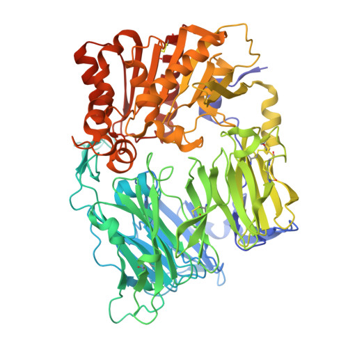
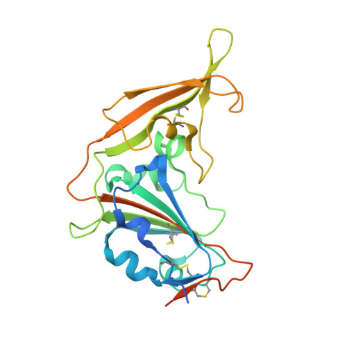
Molecular basis for receptor recognition and broad host tropism for merbecovirus MjHKU4r-CoV-1.

Zhao, Z.Li, X.Chai, Y.Liu, Z.Wang, Q.Gao, G.F.

(2024) EMBO Rep 25: 3116-3136

  • DOI: https://doi.org/10.1038/s44319-024-00169-8
  • Primary Citation of Related Structures:  
    8WKU

  • PubMed Abstract: 

    A novel pangolin-origin MERS-like coronavirus (CoV), MjHKU4r-CoV-1, was recently identified. It is closely related to bat HKU4-CoV, and is infectious in human organs and transgenic mice. MjHKU4r-CoV-1 uses the dipeptidyl peptidase 4 (DPP4 or CD26) receptor for virus entry and has a broad host tropism. However, the molecular mechanism of its receptor binding and determinants of host range are not yet clear. Herein, we determine the structure of the MjHKU4r-CoV-1 spike (S) protein receptor-binding domain (RBD) complexed with human CD26 (hCD26) to reveal the basis for its receptor binding. Measuring binding capacity toward multiple animal receptors for MjHKU4r-CoV-1, mutagenesis analyses, and homology modeling highlight that residue sites 291, 292, 294, 295, 336, and 344 of CD26 are the crucial host range determinants for MjHKU4r-CoV-1. These results broaden our understanding of this potentially high-risk virus and will help us prepare for possible outbreaks in the future.


  • Organizational Affiliation

    CAS Key Laboratory of Pathogen Microbiology and Immunology, Institute of Microbiology, Chinese Academy of Sciences, Beijing, 100101, China.


Macromolecules
Find similar proteins by:  (by identity cutoff)  |  3D Structure
Entity ID: 1
MoleculeChains Sequence LengthOrganismDetailsImage
Dipeptidyl peptidase 4 soluble form734Homo sapiensMutation(s): 0 
Gene Names: DPP4ADCP2CD26
UniProt & NIH Common Fund Data Resources
Find proteins for P27487 (Homo sapiens)
Explore P27487 
Go to UniProtKB:  P27487
PHAROS:  P27487
GTEx:  ENSG00000197635 
Entity Groups  
Sequence Clusters30% Identity50% Identity70% Identity90% Identity95% Identity100% Identity
UniProt GroupP27487
Glycosylation
Glycosylation Sites: 8Go to GlyGen: P27487-1
Sequence Annotations
Expand
  • Reference Sequence
Find similar proteins by:  (by identity cutoff)  |  3D Structure
Entity ID: 2
MoleculeChains Sequence LengthOrganismDetailsImage
MjHKU4r-CoV-1 spike RBD246Manis javanicaMutation(s): 0 
UniProt
Find proteins for A0AAE8ZFM2 (Pangolin coronavirus HKU4/P251T/pangolin/2018)
Explore A0AAE8ZFM2 
Go to UniProtKB:  A0AAE8ZFM2
Entity Groups  
Sequence Clusters30% Identity50% Identity70% Identity90% Identity95% Identity100% Identity
UniProt GroupA0AAE8ZFM2
Glycosylation
Glycosylation Sites: 2
Sequence Annotations
Expand
  • Reference Sequence
Oligosaccharides

Help

Entity ID: 3
MoleculeChains Length2D Diagram Glycosylation3D Interactions
beta-D-mannopyranose-(1-4)-2-acetamido-2-deoxy-beta-D-glucopyranose-(1-4)-[alpha-L-fucopyranose-(1-6)]2-acetamido-2-deoxy-beta-D-glucopyranose
C
4N-Glycosylation
Glycosylation Resources
GlyTouCan:  G32152BH
GlyCosmos:  G32152BH
GlyGen:  G32152BH
Entity ID: 4
MoleculeChains Length2D Diagram Glycosylation3D Interactions
alpha-D-mannopyranose-(1-3)-[alpha-D-mannopyranose-(1-6)]beta-D-mannopyranose-(1-4)-2-acetamido-2-deoxy-beta-D-glucopyranose-(1-4)-[alpha-L-fucopyranose-(1-6)]2-acetamido-2-deoxy-beta-D-glucopyranose
D
6N-Glycosylation
Glycosylation Resources
GlyTouCan:  G82348BZ
GlyCosmos:  G82348BZ
GlyGen:  G82348BZ
Entity ID: 5
MoleculeChains Length2D Diagram Glycosylation3D Interactions
alpha-D-mannopyranose-(1-3)-[alpha-D-mannopyranose-(1-6)]beta-D-mannopyranose-(1-4)-2-acetamido-2-deoxy-beta-D-glucopyranose-(1-4)-2-acetamido-2-deoxy-beta-D-glucopyranose
E, H
5N-Glycosylation
Glycosylation Resources
GlyTouCan:  G22768VO
GlyCosmos:  G22768VO
GlyGen:  G22768VO
Entity ID: 6
MoleculeChains Length2D Diagram Glycosylation3D Interactions
alpha-D-mannopyranose-(1-2)-alpha-D-mannopyranose-(1-2)-alpha-D-mannopyranose-(1-3)-[alpha-D-mannopyranose-(1-6)-alpha-D-mannopyranose-(1-6)]beta-D-mannopyranose-(1-4)-2-acetamido-2-deoxy-beta-D-glucopyranose-(1-4)-2-acetamido-2-deoxy-beta-D-glucopyranose
F
8N-Glycosylation
Glycosylation Resources
GlyTouCan:  G81980VO
GlyCosmos:  G81980VO
GlyGen:  G81980VO
Entity ID: 7
MoleculeChains Length2D Diagram Glycosylation3D Interactions
alpha-D-mannopyranose-(1-6)-beta-D-mannopyranose-(1-4)-2-acetamido-2-deoxy-beta-D-glucopyranose-(1-4)-[alpha-L-fucopyranose-(1-6)]2-acetamido-2-deoxy-beta-D-glucopyranose
G
5N-Glycosylation
Glycosylation Resources
GlyTouCan:  G00395TQ
GlyCosmos:  G00395TQ
GlyGen:  G00395TQ
Entity ID: 8
MoleculeChains Length2D Diagram Glycosylation3D Interactions
beta-D-mannopyranose-(1-4)-2-acetamido-2-deoxy-beta-D-glucopyranose-(1-4)-2-acetamido-2-deoxy-beta-D-glucopyranose
I
3N-Glycosylation
Glycosylation Resources
GlyTouCan:  G15407YE
GlyCosmos:  G15407YE
GlyGen:  G15407YE
Experimental Data & Validation

Experimental Data

  • Method: X-RAY DIFFRACTION
  • Resolution: 3.50 Å
  • R-Value Free: 0.237 
  • R-Value Work: 0.211 
  • R-Value Observed: 0.229 
  • Space Group: P 32 2 1
Unit Cell:
Length ( Å )Angle ( ˚ )
a = 127.501α = 90
b = 127.501β = 90
c = 245.633γ = 120
Software Package:
Software NamePurpose
PHENIXrefinement
XDSdata reduction
XDSdata scaling
PHASERphasing

Structure Validation

View Full Validation Report


Ligand Structure Quality Assessment 


Entry History & Funding Information

Deposition Data


Funding OrganizationLocationGrant Number
National Natural Science Foundation of China (NSFC)China--

Revision History  (Full details and data files)

  • Version 1.0: 2024-06-26
    Type: Initial release
  • Version 1.1: 2024-07-24
    Changes: Database references