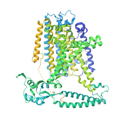
8WG4

mouse TMEM63b in DDM-CHS micelle with YN9303-24 Fab


Experimental Data Snapshot

  • Method: ELECTRON MICROSCOPY
  • Resolution: 3.50 Å
  • Aggregation State: 2D ARRAY 
  • Reconstruction Method: SINGLE PARTICLE 

Starting Model: in silico
View more details

wwPDB Validation   3D Report Full Report


This is version 1.0 of the entry. See complete history


Literature

Mechanosensitive channel TMEM63B functions as a plasma membrane lipid scramblase

Miyata, Y.Takahashi, K.Lee, Y.Sultan, C.S.Kuribayashi, R.Takahashi, M.Hata, K.Bamba, T.Izumi, Y.Liu, K.Uemura, T.Nomura, N.Iwata, S.Nagata, S.Nishizawa, T.Segawa, K.

To be published.

Macromolecules
Find similar proteins by:  (by identity cutoff)  |  3D Structure
Entity ID: 1
MoleculeChains Sequence LengthOrganismDetailsImage
CSC1-like protein 2,Green fluorescent protein1,117Mus musculusAequorea victoria
This entity is chimeric
Mutation(s): 4 
Gene Names: Tmem63bGFP
Membrane Entity: Yes 
UniProt & NIH Common Fund Data Resources
Find proteins for P42212 (Aequorea victoria)
Explore P42212 
Go to UniProtKB:  P42212
Find proteins for Q3TWI9 (Mus musculus)
Explore Q3TWI9 
Go to UniProtKB:  Q3TWI9
IMPC:  MGI:2387609
Entity Groups  
Sequence Clusters30% Identity50% Identity70% Identity90% Identity95% Identity100% Identity
UniProt GroupsQ3TWI9P42212
Sequence Annotations
Expand
  • Reference Sequence
Small Molecules
Ligands 2 Unique
IDChains Name / Formula / InChI Key2D Diagram3D Interactions
LBN
Query on LBN

Download Ideal Coordinates CCD File 
B [auth A]1-palmitoyl-2-oleoyl-sn-glycero-3-phosphocholine
C42 H82 N O8 P
WTJKGGKOPKCXLL-VYOBOKEXSA-N
Y01
Query on Y01

Download Ideal Coordinates CCD File 
C [auth A]
D [auth A]
E [auth A]
F [auth A]
G [auth A]
C [auth A],
D [auth A],
E [auth A],
F [auth A],
G [auth A],
H [auth A],
I [auth A]
CHOLESTEROL HEMISUCCINATE
C31 H50 O4
WLNARFZDISHUGS-MIXBDBMTSA-N
Modified Residues  1 Unique
IDChains TypeFormula2D DiagramParent
P1L
Query on P1L
A
L-PEPTIDE LINKINGC19 H37 N O3 SCYS
Experimental Data & Validation

Experimental Data

  • Method: ELECTRON MICROSCOPY
  • Resolution: 3.50 Å
  • Aggregation State: 2D ARRAY 
  • Reconstruction Method: SINGLE PARTICLE 
EM Software:
TaskSoftware PackageVersion
MODEL REFINEMENTServalcat0.2.24
RECONSTRUCTIONcryoSPARC4.2.1

Structure Validation

View Full Validation Report


Entry History & Funding Information

Deposition Data


Funding OrganizationLocationGrant Number
Japan Society for the Promotion of Science (JSPS)Japan--

Revision History  (Full details and data files)

  • Version 1.0: 2024-09-25
    Type: Initial release