The ultra-high affinity transport proteins of ubiquitous marine bacteria.
Clifton, B.E., Alcolombri, U., Uechi, G.I., Jackson, C.J., Laurino, P.(2024) Nature 634: 721-728
- PubMed: 39261732 Search on PubMedSearch on PubMed Central
- DOI: https://doi.org/10.1038/s41586-024-07924-w
- Primary Citation Related Structures:
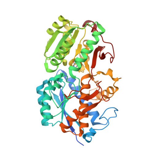
8HQQ, 8HQR, 8KD0, 8WCH - PubMed Abstract:
SAR11 bacteria are the most abundant microorganisms in the surface ocean 1 and have global biogeochemical importance 2-4 . To thrive in their competitive oligotrophic environment, these bacteria rely heavily on solute-binding proteins that facilitate uptake of specific substrates via membrane transporters 5,6 . The functions and properties of these transport proteins are key factors in the assimilation of dissolved organic matter and biogeochemical cycling of nutrients in the ocean, but they have remained largely inaccessible to experimental investigation. Here we performed genome-wide experimental characterization of all solute-binding proteins in a prototypical SAR11 bacterium, revealing specific functions and general trends in their properties that contribute to the success of SAR11 bacteria in oligotrophic environments. We found that the solute-binding proteins of SAR11 bacteria have extremely high binding affinity (dissociation constant >20 pM) and high binding specificity, revealing molecular mechanisms of oligotrophic adaptation. Our functional data have uncovered new carbon sources for SAR11 bacteria and enable accurate biogeographical analysis of SAR11 substrate uptake capabilities throughout the ocean. This study provides a comprehensive view of the substrate uptake capabilities of ubiquitous marine bacteria, providing a necessary foundation for understanding their contribution to assimilation of dissolved organic matter in marine ecosystems.
- Protein Engineering and Evolution Unit, Okinawa Institute of Science and Technology Graduate University, Onna, Japan. benjamin.clifton@oist.jp.
Organizational Affiliation: