Targeting DNA junction sites by bis-intercalators induces topological changes with potent antitumor effects.
Huang, S.C., Chen, C.W., Satange, R., Hsieh, C.C., Chang, C.C., Wang, S.C., Peng, C.L., Chen, T.L., Chiang, M.H., Horng, Y.C., Hou, M.H.(2024) Nucleic Acids Res 52: 9303-9316
- PubMed: 39036959 Search on PubMed
- DOI: https://doi.org/10.1093/nar/gkae643
- Primary Citation Related Structures:
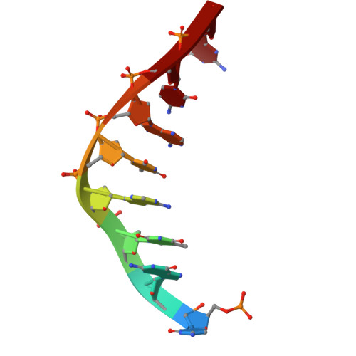
8W76, 8W7W, 8WQ7 - PubMed Abstract:
Targeting inter-duplex junctions in catenated DNA with bidirectional bis-intercalators is a potential strategy for enhancing anticancer effects. In this study, we used d(CGTATACG)2, which forms a tetraplex base-pair junction that resembles the DNA-DNA contact structure, as a model target for two alkyl-linked diaminoacridine bis-intercalators, DA4 and DA5. Cross-linking of the junction site by the bis-intercalators induced substantial structural changes in the DNA, transforming it from a B-form helical end-to-end junction to an over-wounded side-by-side inter-duplex conformation with A-DNA characteristics and curvature. These structural perturbations facilitated the angled intercalation of DA4 and DA5 with propeller geometry into two adjacent duplexes. The addition of a single carbon to the DA5 linker caused a bend that aligned its chromophores with CpG sites, enabling continuous stacking and specific water-mediated interactions at the inter-duplex contacts. Furthermore, we have shown that the different topological changes induced by DA4 and DA5 lead to the inhibition of topoisomerase 2 activities, which may account for their antitumor effects. Thus, this study lays the foundations for bis-intercalators targeting biologically relevant DNA-DNA contact structures for anticancer drug development.
- Doctoral Program in Medical Biotechnology, National Chung Hsing University, Taichung 402, Taiwan.
Organizational Affiliation: