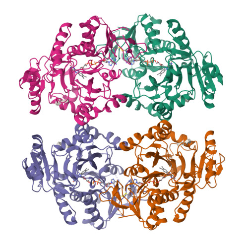
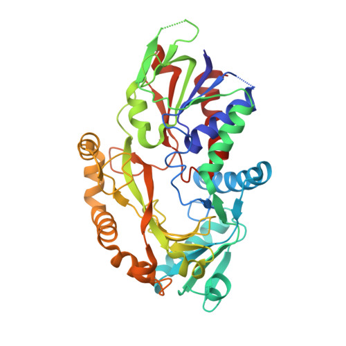
8W75

Structure of Drosophila melanogaster L-2-hydroxyglutarate dehydrogenase


Experimental Data Snapshot

  • Method: X-RAY DIFFRACTION
  • Resolution: 2.85 Å
  • R-Value Free: 0.294 
  • R-Value Work: 0.243 
  • R-Value Observed: 0.246 

Starting Model: in silico
View more details

wwPDB Validation   3D Report Full Report


Ligand Structure Quality Assessment 


This is version 1.3 of the entry. See complete history


Literature

Structure and biochemical characterization of l-2-hydroxyglutarate dehydrogenase and its role in the pathogenesis of l-2-hydroxyglutaric aciduria.

Yang, J.Chen, X.Jin, S.Ding, J.

(2023) J Biol Chem 300: 105491-105491

  • DOI: https://doi.org/10.1016/j.jbc.2023.105491
  • Primary Citation of Related Structures:  
    8W75, 8W78, 8W7F

  • PubMed Abstract: 

    l-2-hydroxyglutarate dehydrogenase (L2HGDH) is a mitochondrial membrane-associated metabolic enzyme, which catalyzes the oxidation of l-2-hydroxyglutarate (l-2-HG) to 2-oxoglutarate (2-OG). Mutations in human L2HGDH lead to abnormal accumulation of l-2-HG, which causes a neurometabolic disorder named l-2-hydroxyglutaric aciduria (l-2-HGA). Here, we report the crystal structures of Drosophila melanogaster L2HGDH (dmL2HGDH) in FAD-bound form and in complex with FAD and 2-OG and show that dmL2HGDH exhibits high activity and substrate specificity for l-2-HG. dmL2HGDH consists of an FAD-binding domain and a substrate-binding domain, and the active site is located at the interface of the two domains with 2-OG binding to the re-face of the isoalloxazine moiety of FAD. Mutagenesis and activity assay confirmed the functional roles of key residues involved in the substrate binding and catalytic reaction and showed that most of the mutations of dmL2HGDH equivalent to l-2-HGA-associated mutations of human L2HGDH led to complete loss of the activity. The structural and biochemical data together reveal the molecular basis for the substrate specificity and catalytic mechanism of L2HGDH and provide insights into the functional roles of human L2HGDH mutations in the pathogeneses of l-2-HGA.


  • Organizational Affiliation

    State Key Laboratory of Molecular Biology, Shanghai Institute of Biochemistry and Cell Biology, Center for Excellence in Molecular Cell Science, Chinese Academy of Sciences, University of Chinese Academy of Sciences, Shanghai, China.


Macromolecules
Find similar proteins by:  (by identity cutoff)  |  3D Structure
Entity ID: 1
MoleculeChains Sequence LengthOrganismDetailsImage
FI05204pA [auth D],
B [auth A],
C [auth B],
D [auth C]
415Drosophila melanogasterMutation(s): 0 
Gene Names: L2HGDHCG10639-RAdL2HGDHDmel\CG10639L-2-HGDHCG10639Dmel_CG10639
EC: 1.1.3.15 (PDB Primary Data), 1.1.99.2 (PDB Primary Data)
UniProt
Find proteins for Q9VJ28 (Drosophila melanogaster)
Explore Q9VJ28 
Go to UniProtKB:  Q9VJ28
Entity Groups  
Sequence Clusters30% Identity50% Identity70% Identity90% Identity95% Identity100% Identity
UniProt GroupQ9VJ28
Sequence Annotations
Expand
  • Reference Sequence
Small Molecules
Ligands 2 Unique
IDChains Name / Formula / InChI Key2D Diagram3D Interactions
FAD (Subject of Investigation/LOI)
Query on FAD

Download Ideal Coordinates CCD File 
E [auth D],
G [auth A],
I [auth B],
K [auth C]
FLAVIN-ADENINE DINUCLEOTIDE
C27 H33 N9 O15 P2
VWWQXMAJTJZDQX-UYBVJOGSSA-N
LMT
Query on LMT

Download Ideal Coordinates CCD File 
F [auth D],
H [auth A],
J [auth B],
L [auth C]
DODECYL-BETA-D-MALTOSIDE
C24 H46 O11
NLEBIOOXCVAHBD-QKMCSOCLSA-N
Experimental Data & Validation

Experimental Data

  • Method: X-RAY DIFFRACTION
  • Resolution: 2.85 Å
  • R-Value Free: 0.294 
  • R-Value Work: 0.243 
  • R-Value Observed: 0.246 
  • Space Group: P 1 21 1
Unit Cell:
Length ( Å )Angle ( ˚ )
a = 100.115α = 90
b = 103.178β = 108.61
c = 102.538γ = 90
Software Package:
Software NamePurpose
PHENIXrefinement
HKL-2000data reduction
HKL-2000data scaling
PHENIXphasing

Structure Validation

View Full Validation Report


Ligand Structure Quality Assessment 


Entry History & Funding Information

Deposition Data


Funding OrganizationLocationGrant Number
Chinese Academy of SciencesChinaXDB37030305
National Natural Science Foundation of China (NSFC)China32071190

Revision History  (Full details and data files)

  • Version 1.0: 2023-11-29
    Type: Initial release
  • Version 1.1: 2023-12-06
    Changes: Database references
  • Version 1.2: 2023-12-20
    Changes: Database references
  • Version 1.3: 2024-11-13
    Changes: Structure summary