8W74

Fe-O nanocluster of form-II in the 4-fold channel of Ureaplasma diversum ferritin


Experimental Data Snapshot

  • Method: X-RAY DIFFRACTION
  • Resolution: 2.50 Å
  • R-Value Free: 0.236 
  • R-Value Work: 0.183 
  • R-Value Observed: 0.188 

Starting Model: experimental
View more details

wwPDB Validation   3D Report Full Report


This is version 1.0 of the entry. See complete history


Literature

Growth Process of Fe-O Nanoclusters with Different Sizes Biosynthesized by Protein Nanocages.

Wang, W.Xi, H.Fu, D.Ma, D.Gong, W.Zhao, Y.Li, X.Wu, L.Guo, Y.Zhao, G.Wang, H.

(2024) J Am Chem Soc 146: 11657-11668

  • DOI: https://doi.org/10.1021/jacs.3c13830
  • Primary Citation of Related Structures:  
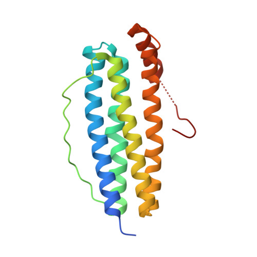
    8W6M, 8W6Q, 8W6S, 8W6U, 8W6Y, 8W73, 8W74, 8W79, 8W7B, 8W7O, 8W7Q, 8W7T, 8W7U, 8W7V, 8WPT, 8WPV, 8WQU, 8WQV, 8WQX, 8WQY, 8WR0

  • PubMed Abstract: 

    All protein-directed syntheses of metal nanoclusters (NCs) and nanoparticles (NPs) have attracted considerable attention because protein scaffolds provide a unique metal coordination environment and can adjust the shape and morphology of NCs and NPs. However, the detailed formation mechanisms of NCs or NPs directed by protein templates remain unclear. In this study, by taking advantage of the ferritin nanocage as a biotemplate to monitor the growth of Fe-O NCs as a function of time, we synthesized a series of iron NCs with different sizes and shapes and subsequently solved their corresponding three-dimensional atomic-scale structures by X-ray protein crystallography and cryo-electron microscopy. The time-dependent structure analyses revealed the growth process of these Fe-O NCs with the 4-fold channel of ferritin as nucleation sites. To our knowledge, the newly biosynthesized Fe 35 O 23 Glu 12 represents the largest Fe-O NCs with a definite atomic structure. This study contributes to our understanding of the formation mechanism of iron NCs and provides an effective method for metal NC synthesis.


  • Organizational Affiliation

    Key Laboratory of Chemical Biology and Molecular Engineering of the Education Ministry, Institute of Molecular Science, Shanxi University, Taiyuan 030006, China.


Macromolecules
Find similar proteins by:  (by identity cutoff)  |  3D Structure
Entity ID: 1
MoleculeChains Sequence LengthOrganismDetailsImage
Ferritin181Ureaplasma diversumMutation(s): 0 
Gene Names: UDIV_5880
UniProt
Find proteins for A0A084EX36 (Ureaplasma diversum NCTC 246)
Explore A0A084EX36 
Go to UniProtKB:  A0A084EX36
Entity Groups  
Sequence Clusters30% Identity50% Identity70% Identity90% Identity95% Identity100% Identity
UniProt GroupA0A084EX36
Sequence Annotations
Expand
  • Reference Sequence
Small Molecules
Ligands 1 Unique
IDChains Name / Formula / InChI Key2D Diagram3D Interactions
FE (Subject of Investigation/LOI)
Query on FE

Download Ideal Coordinates CCD File 
B [auth A]
C [auth A]
D [auth A]
E [auth A]
F [auth A]
B [auth A],
C [auth A],
D [auth A],
E [auth A],
F [auth A],
G [auth A],
H [auth A],
I [auth A],
J [auth A],
K [auth A],
L [auth A]
FE (III) ION
Fe
VTLYFUHAOXGGBS-UHFFFAOYSA-N
Experimental Data & Validation

Experimental Data

  • Method: X-RAY DIFFRACTION
  • Resolution: 2.50 Å
  • R-Value Free: 0.236 
  • R-Value Work: 0.183 
  • R-Value Observed: 0.188 
  • Space Group: F 4 3 2
Unit Cell:
Length ( Å )Angle ( ˚ )
a = 179.646α = 90
b = 179.646β = 90
c = 179.646γ = 90
Software Package:
Software NamePurpose
PHENIXrefinement
HKL-2000data scaling
HKL-2000data reduction
PHASERphasing

Structure Validation

View Full Validation Report


Entry History & Funding Information

Deposition Data


Funding OrganizationLocationGrant Number
National Natural Science Foundation of China (NSFC)China62075118, 21601112

Revision History  (Full details and data files)

  • Version 1.0: 2024-06-12
    Type: Initial release