8W6Z | pdb_00008w6z

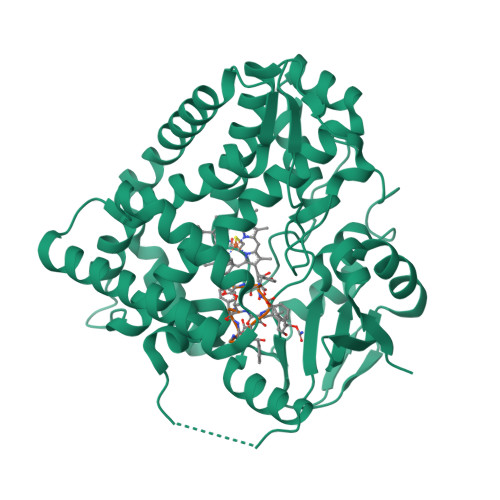
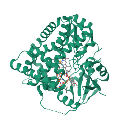
Substrate-bound crystal structure of a P450 enzyme DmlH that catalyze intramolecular phenol coupling in the biosynthesis of cihanmycins


Experimental Data Snapshot

  • Method: X-RAY DIFFRACTION
  • Resolution: 2.20 Å
  • R-Value Free: 
    0.234 (Depositor), 0.235 (DCC) 
  • R-Value Work: 
    0.183 (Depositor), 0.182 (DCC) 
  • R-Value Observed: 
    0.185 (Depositor) 

Starting Model: experimental
View more details

wwPDB Validation   3D Report Full Report


Ligand Structure Quality Assessment 

Created with Raphaël 2.3.0Worse 01 BetterLigand structure goodness of fit to experimental dataBest fitted R8LClick on this verticalbar to view details

This is version 2.1 of the entry. See complete history


Literature

Discovery and Biosynthesis of Cihanmycins Reveal Cytochrome P450-Catalyzed Intramolecular C-O Phenol Coupling Reactions.

Fang, C.Zhang, L.Wang, Y.Xiong, W.Yan, Z.Zhang, W.Zhang, Q.Wang, B.Zhu, Y.Zhang, C.

(2024) J Am Chem Soc 

  • DOI: https://doi.org/10.1021/jacs.4c02841
  • Primary Citation of Related Structures:  
    8W6Z, 8W72

  • PubMed Abstract: 

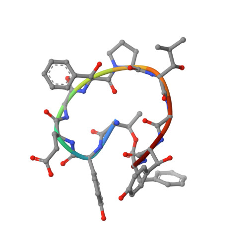
    Cinnamoyl-containing nonribosomal peptides (CCNPs) constitute a unique family of natural products. The enzyme mechanism for the biaryl phenol coupling reaction of the bicyclic CCNPs remains unclear. Herein, we report the discovery of two new arabinofuranosylated bicyclic CCNPs cihanmycins (CHMs) A ( 1 ) and B ( 2 ) from Amycolatopsis cihanbeyliensis DSM 45679 and the identification of the CHM biosynthetic gene cluster ( cih BGC) by heterologous expression in Streptomyces lividans SBT18 to afford CHMs C ( 3 ) and D ( 4 ). The structure of 1 was confirmed by X-ray diffraction analysis. Three cytochrome P450 enzyme (CYP)-encoding genes cih26 , cih32 , and cih33 were individually inactivated in the heterologous host to produce CHMs E ( 5 ), F ( 6 ), and G ( 7 ), respectively. The structures of 5 and 6 indicated that Cih26 was responsible for the hydroxylation and epoxidation of the cinnamoyl moiety, and Cih32 should catalyze the β-hydroxylation of three amino acid residues. Cih33 and its homologues DmlH and EpcH were biochemically verified to convert CHM G ( 7 ) with a monocyclic structure to a bicyclic skeleton of CHM C ( 3 ) through an intramolecular C-O phenol coupling reaction. The substrate 7 -bound crystal structure of DmlH not only established the structure of 7 , which was difficult for NMR analysis for displaying anomalous splitting signals, but also provided the binding mode of macrocyclic peptides recognized by these intramolecular C-O coupling CYPs. In addition, computational studies revealed a water-mediated diradical mechanism for the C-O phenol coupling reaction. These findings have shed important mechanistic insights into the CYP-catalyzed phenol coupling reactions.


  • Organizational Affiliation

    Key Laboratory of Tropical Marine Bio-resources and Ecology, Guangdong Key Laboratory of Marine Materia Medica, South China Sea Institute of Oceanology, Chinese Academy of Sciences, 164 West Xingang Road, Guangzhou 510301, China.


Macromolecules
Find similar proteins by:  (by identity cutoff)  |  3D Structure
Entity ID: 1
MoleculeChains Sequence LengthOrganismDetailsImage
Cytochrome P450428Streptomyces sp.Mutation(s): 0 
Entity Groups  
Sequence Clusters30% Identity50% Identity70% Identity90% Identity95% Identity100% Identity
Sequence Annotations
Expand
  • Reference Sequence

Find similar proteins by:  Sequence   |   3D Structure  

Entity ID: 2
MoleculeChains Sequence LengthOrganismDetailsImage
THR-D4P-GLU-GLY-BB8-PRO-KJW-GLY-A1D5P-DPN10Streptomyces sp.Mutation(s): 0 
Entity Groups  
Sequence Clusters30% Identity50% Identity70% Identity90% Identity95% Identity100% Identity
Sequence Annotations
Expand
  • Reference Sequence
Small Molecules
Ligands 2 Unique
IDChains Name / Formula / InChI Key2D Diagram3D Interactions
HEM
Query on HEM

Download Ideal Coordinates CCD File 
C [auth A]PROTOPORPHYRIN IX CONTAINING FE
C34 H32 Fe N4 O4
KABFMIBPWCXCRK-RGGAHWMASA-L
R8L (Subject of Investigation/LOI)
Query on R8L

Download Ideal Coordinates CCD File 
D [auth B](E)-3-[2-[(2R,3S)-3-[(1R)-1-aminocarbonyloxypropyl]oxiran-2-yl]phenyl]prop-2-enoic acid
C15 H17 N O5
APIHDLCQFHISLU-PQPVHHHVSA-N
Modified Residues  2 Unique
IDChains TypeFormula2D DiagramParent
A1D5P
Query on A1D5P
B
D-PEPTIDE LINKINGC9 H11 N O4TYR
BB8
Query on BB8
B
L-PEPTIDE LINKINGC9 H11 N O3PHE
Biologically Interesting Molecules (External Reference) 1 Unique
Experimental Data & Validation

Experimental Data

  • Method: X-RAY DIFFRACTION
  • Resolution: 2.20 Å
  • R-Value Free:  0.234 (Depositor), 0.235 (DCC) 
  • R-Value Work:  0.183 (Depositor), 0.182 (DCC) 
  • R-Value Observed: 0.185 (Depositor) 
Space Group: P 21 21 21
Unit Cell:
Length ( Å )Angle ( ˚ )
a = 36.082α = 90
b = 100.162β = 90
c = 105.811γ = 90
Software Package:
Software NamePurpose
PHENIXrefinement
Aimlessdata scaling
MOLREPphasing
CrysalisProdata reduction

Structure Validation

View Full Validation Report


Ligand Structure Quality Assessment 

Created with Raphaël 2.3.0Worse 01 BetterLigand structure goodness of fit to experimental dataBest fitted R8LClick on this verticalbar to view details

Entry History & Funding Information

Deposition Data


Funding OrganizationLocationGrant Number
National Natural Science Foundation of China (NSFC)China22177118
National Natural Science Foundation of China (NSFC)China31820103003
National Natural Science Foundation of China (NSFC)China31630004

Revision History  (Full details and data files)

  • Version 1.0: 2024-06-12
    Type: Initial release
  • Version 2.0: 2024-06-19
    Changes: Atomic model, Data collection, Derived calculations, Structure summary
  • Version 2.1: 2024-12-25
    Changes: Database references, Structure summary