8VYJ | pdb_00008vyj

Structure of full-length human cardiac sodium channel - Class-I.


Experimental Data Snapshot

  • Method: ELECTRON MICROSCOPY
  • Resolution: 3.60 Å
  • Aggregation State: PARTICLE 
  • Reconstruction Method: SINGLE PARTICLE 

wwPDB Validation 3D Report Full Report

Validation slider image for 8VYJ

This is version 1.1 of the entry. See complete history

Literature

Structural basis of human Na v 1.5 gating mechanisms.

Biswas, R.Lopez-Serrano, A.L.Purohit, A.Ramirez-Navarro, A.Huang, H.L.Grandinetti, G.Cheng, X.Heissler, S.M.Deschenes, I.Chinthalapudi, K.

(2025) Proc Natl Acad Sci U S A 122: e2416181122-e2416181122

  • DOI: https://doi.org/10.1073/pnas.2416181122
  • Primary Citation Related Structures: 
    8VYJ, 8VYK

  • PubMed Abstract: 

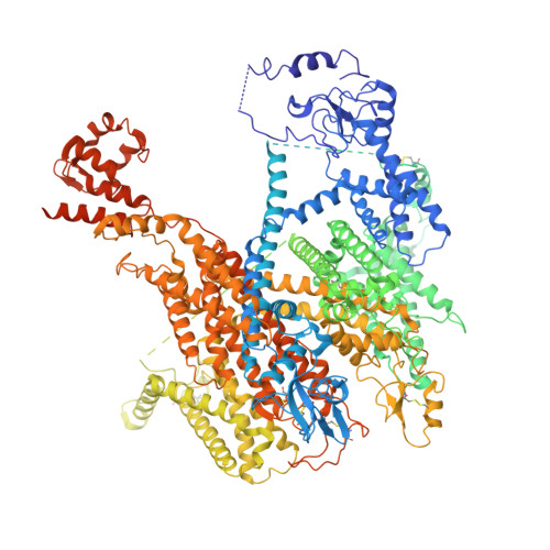
    Voltage-gated Na v 1.5 channels are central to the generation and propagation of cardiac action potentials. Aberrations in their function are associated with a wide spectrum of cardiac diseases including arrhythmias and heart failure. Despite decades of progress in Na v 1.5 biology, the lack of structural insights into intracellular regions has hampered our understanding of its gating mechanisms. Here, we present two cryo-EM structures of human Na v 1.5 in open states, revealing sequential conformational changes in gating charges of the voltage-sensing domains (VSDs) and several intracellular regions. Despite the channel being in the open state, these structures show repositioning, but no dislodging of the IFM motif in the receptor site. Molecular dynamics analyses show our structures with CTD conduct Na + ions. Notably, our structural findings highlight a dynamic C-terminal domain (CTD) and III-IV linker interaction, which regulates the conformation of VSDs and pore opening. Electrophysiological studies confirm that disrupting this interaction alters fast inactivation of Na v 1.5. Together, our structure-function studies establish a foundation for understanding the gating mechanisms of Na v 1.5 and the mechanisms underlying CTD-related channelopathies.


  • Organizational Affiliation
    • Department of Physiology and Cell Biology, Dorothy M. Davis Heart and Lung Research Institute, College of Medicine, The Ohio State University, Columbus, OH 43210.

Macromolecule Content 

  • Total Structure Weight: 234.97 kDa 
  • Atom Count: 11,715 
  • Modeled Residue Count: 1,395 
  • Deposited Residue Count: 2,016 
  • Unique protein chains: 1

Macromolecules

Find similar proteins by:|  3D Structure
Entity ID: 1
MoleculeChains  Sequence LengthOrganismDetailsImage
Sodium channel protein type 5 subunit alpha2,016Homo sapiensMutation(s): 0 
Gene Names: SCN5A
Membrane Entity: Yes 
UniProt & NIH Common Fund Data Resources
Find proteins for Q14524 (Homo sapiens)
Explore Q14524 
Go to UniProtKB:  Q14524
PHAROS:  Q14524
GTEx:  ENSG00000183873 
Entity Groups
Sequence Clusters30% Identity50% Identity70% Identity90% Identity95% Identity100% Identity
UniProt GroupQ14524
Glycosylation
Glycosylation Sites: 4Go to GlyGen: Q14524-1
Sequence Annotations
Expand
Reference Sequence

Oligosaccharides

Help  
Entity ID: 2
MoleculeChains Length2D Diagram GlycosylationD Interactions
2-acetamido-2-deoxy-beta-D-glucopyranose-(1-4)-2-acetamido-2-deoxy-beta-D-glucopyranose-(1-4)-2-acetamido-2-deoxy-beta-D-glucopyranose-(1-4)-2-acetamido-2-deoxy-beta-D-glucopyranose-(1-4)-2-acetamido-2-deoxy-beta-D-glucopyranose
B
5N/A
Glycosylation Resources
GlyTouCan: G07670VS
GlyCosmos: G07670VS
GlyGen: G07670VS
Entity ID: 3
MoleculeChains Length2D Diagram GlycosylationD Interactions
2-acetamido-2-deoxy-beta-D-glucopyranose-(1-4)-2-acetamido-2-deoxy-beta-D-glucopyranose
C, D, E
2N-Glycosylation
Glycosylation Resources
GlyTouCan: G42666HT
GlyCosmos: G42666HT
GlyGen: G42666HT

Small Molecules

Ligands 3 Unique
IDChains Name / Formula / InChI Key2D Diagram3D Interactions
6OU
(Subject of Investigation/LOI)

Query on 6OU


Download:Ideal Coordinates CCD File
F [auth A],
Q [auth A]
[(2~{R})-1-[2-azanylethoxy(oxidanyl)phosphoryl]oxy-3-hexadecanoyloxy-propan-2-yl] (~{Z})-octadec-9-enoate
C39 H76 N O8 P
FHQVHHIBKUMWTI-OTMQOFQLSA-N
Y01
(Subject of Investigation/LOI)

Query on Y01


Download:Ideal Coordinates CCD File
G [auth A]
H [auth A]
I [auth A]
J [auth A]
K [auth A]
G [auth A],
H [auth A],
I [auth A],
J [auth A],
K [auth A],
L [auth A],
M [auth A]
CHOLESTEROL HEMISUCCINATE
C31 H50 O4
WLNARFZDISHUGS-MIXBDBMTSA-N
NAG
(Subject of Investigation/LOI)

Query on NAG


Download:Ideal Coordinates CCD File
N [auth A],
O [auth A],
P [auth A]
2-acetamido-2-deoxy-beta-D-glucopyranose
C8 H15 N O6
OVRNDRQMDRJTHS-FMDGEEDCSA-N

Experimental Data & Validation

Experimental Data

  • Method: ELECTRON MICROSCOPY
  • Resolution: 3.60 Å
  • Aggregation State: PARTICLE 
  • Reconstruction Method: SINGLE PARTICLE 
EM Software:
TaskSoftware PackageVersion
MODEL REFINEMENTPHENIX
RECONSTRUCTIONcryoSPARC

Structure Validation

View Full Validation Report


Entry History 

& Funding Information

Deposition Data


Funding OrganizationLocationGrant Number
National Institutes of Health/National Heart, Lung, and Blood Institute (NIH/NHLBI)United StatesR01HL094450

Revision History  (Full details and data files)

  • Version 1.0: 2025-02-12
    Type: Initial release
  • Version 1.1: 2026-02-25
    Changes: Data collection, Database references