


Crystal Structure Analysis of PARP1 in complex with a compound
Seo, H.-S., Dhe-Paganon, S., Arthanari, H.To be published.
Experimental Data Snapshot
Starting Model: experimental
View more details
Entity ID: 1 | |||||
|---|---|---|---|---|---|
| Molecule | Chains | Sequence Length | Organism | Details | Image |
| Poly [ADP-ribose] polymerase 1, processed C-terminus | 352 | Homo sapiens | Mutation(s): 0 Gene Names: PARP1, ADPRT, PPOL EC: 2.4.2 (UniProt), 2.4.2.30 (UniProt) | 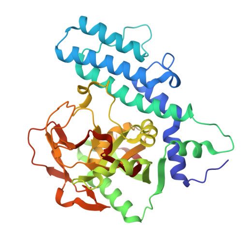 | |
UniProt & NIH Common Fund Data Resources | |||||
PHAROS: P09874 GTEx: ENSG00000143799 | |||||
Entity Groups | |||||
| Sequence Clusters | 30% Identity50% Identity70% Identity90% Identity95% Identity100% Identity | ||||
| UniProt Group | P09874 | ||||
Sequence AnnotationsExpand | |||||
Reference Sequence | |||||
| Ligands 2 Unique | |||||
|---|---|---|---|---|---|
| ID | Chains | Name / Formula / InChI Key | 2D Diagram | 3D Interactions | |
| A1AEO (Subject of Investigation/LOI) Download:Ideal Coordinates CCD File | C [auth A], H [auth B] | (4S)-1-{2-fluoro-5-[(4-oxo-3,4-dihydrophthalazin-1-yl)methyl]benzoyl}-4-hydroxy-L-proline C21 H18 F N3 O5 JKGJSRKNKYMRHL-SGTLLEGYSA-N |  | ||
| SO4 Download:Ideal Coordinates CCD File | D [auth A] E [auth A] F [auth A] G [auth A] I [auth B] | SULFATE ION O4 S QAOWNCQODCNURD-UHFFFAOYSA-L |  | ||
| Length ( Å ) | Angle ( ˚ ) |
|---|---|
| a = 48.2 | α = 90 |
| b = 93.14 | β = 90 |
| c = 165.55 | γ = 90 |
| Software Name | Purpose |
|---|---|
| PHENIX | refinement |
| xia2 | data scaling |
| xia2 | data reduction |
| PHASER | phasing |
| Funding Organization | Location | Grant Number |
|---|---|---|
| National Institutes of Health/National Cancer Institute (NIH/NCI) | United States | -- |


