8VY0 | pdb_00008vy0

Mango II bound to 365A-087


Experimental Data Snapshot

  • Method: X-RAY DIFFRACTION
  • Resolution: 3.10 Å
  • R-Value Free: 
    0.260 (Depositor), 0.260 (DCC) 
  • R-Value Work: 
    0.214 (Depositor), 0.215 (DCC) 
  • R-Value Observed: 
    0.212 (Depositor) 

Starting Model: experimental
View more details

wwPDB Validation 3D Report Full Report

Validation slider image for 8VY0

Ligand Structure Quality Assessment 


This is version 1.1 of the entry. See complete history

Literature

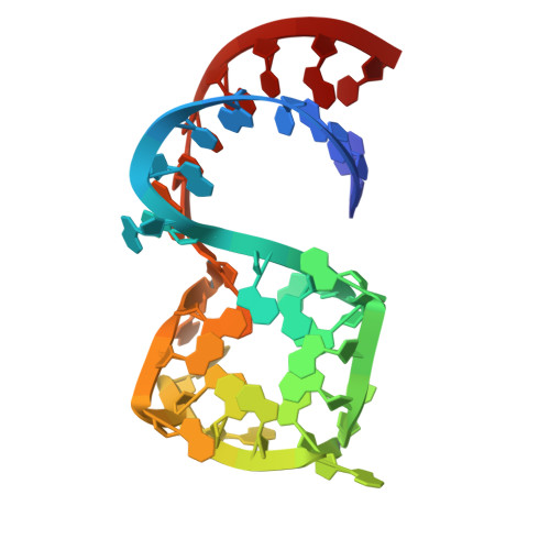
Structure-informed design of an ultrabright RNA-activated fluorophore.

Yang, M.Prestwood, P.R.Passalacqua, L.F.M.Balaratnam, S.Fullenkamp, C.R.Arney, J.W.Weeks, K.M.Ferre-D'Amare, A.Schneekloth Jr., J.S.

(2025) Nat Chem 17: 1188-1195

  • DOI: https://doi.org/10.1038/s41557-025-01832-w
  • Primary Citation Related Structures: 
    8VXX, 8VXZ, 8VY0, 8VY1

  • PubMed Abstract: 

    RNA-based fluorogenic aptamers, such as Mango, are uniquely powerful tools for imaging RNA that activate the fluorescence of a weakly or non-fluorescent small molecule when bound. A central challenge has been to develop brighter, more specific and high-affinity aptamer-ligand systems for cellular imaging. Here we report an ultrabright fluorophore for the Mango II system discovered using a structure-informed, fragment-based small-molecule microarray approach. This dye-termed SALAD1 (structure-informed, array-enabled LigAnD 1)-exhibits subnanomolar aptamer affinity and 3.5-fold brighter fluorescence than Mango II-TO1-biotin pair, a widely used fluorogenic system. Performance was improved by modulating RNA-dye molecular recognition without altering the fluorophore's π-system. High-resolution X-ray structures reveal the binding mode for SALAD1, which exhibits improved pocket occupancy, a more defined binding pose and a unique bonding interaction with potassium. SALAD1 is cell-permeable and facilitates improved in-cell confocal RNA imaging. This work introduces an additional RNA-activated fluorophore demonstrating how fragment-based ligand discovery can be used to create high-performance ligands for RNA targets.


  • Organizational Affiliation
    • Chemical Biology Laboratory, Center for Cancer Research, National Cancer Institute, Frederick, MD, USA.

Macromolecule Content 

  • Total Structure Weight: 37.71 kDa 
  • Atom Count: 2,467 
  • Modeled Residue Count: 108 
  • Deposited Residue Count: 108 
  • Unique nucleic acid chains: 1

Macromolecules

Find similar nucleic acids by:  (by identity cutoff) 
Entity ID: 1
MoleculeChains LengthOrganismImage
Chains: A,B,C
A, B, C
36synthetic construct
Sequence Annotations
Expand
Reference Sequence

Small Molecules

Ligands 2 Unique
IDChains Name / Formula / InChI Key2D Diagram3D Interactions
A1AEE
(Subject of Investigation/LOI)

Query on A1AEE


Download:Ideal Coordinates CCD File
H [auth A],
M [auth B],
P [auth C]
2-[(E)-(1-methylquinolin-4(1H)-ylidene)methyl]-3-[2-oxo-2-({[(2P)-2-(1H-pyrazol-1-yl)phenyl]methyl}amino)ethyl]-1,3-benzothiazol-3-ium
C30 H26 N5 O S
IPCKTFJCCLGQKH-UHFFFAOYSA-O
K

Query on K


Download:Ideal Coordinates CCD File
D [auth A]
E [auth A]
F [auth A]
G [auth A]
I [auth B]
D [auth A],
E [auth A],
F [auth A],
G [auth A],
I [auth B],
J [auth B],
K [auth B],
L [auth B],
N [auth C],
O [auth C]
POTASSIUM ION
K
NPYPAHLBTDXSSS-UHFFFAOYSA-N

Experimental Data & Validation

Experimental Data

  • Method: X-RAY DIFFRACTION
  • Resolution: 3.10 Å
  • R-Value Free:  0.260 (Depositor), 0.260 (DCC) 
  • R-Value Work:  0.214 (Depositor), 0.215 (DCC) 
  • R-Value Observed: 0.212 (Depositor) 
Space Group: C 2 2 21
Unit Cell:
Length ( Å )Angle ( ˚ )
a = 37.254α = 90
b = 184.109β = 90
c = 108.562γ = 90
Software Package:
Software NamePurpose
PHENIXrefinement
PHASERphasing
xia2data reduction
DIALSdata scaling

Structure Validation

View Full Validation Report


Ligand Structure Quality Assessment 


Entry History 

& Funding Information

Deposition Data


Funding OrganizationLocationGrant Number
National Institutes of Health/National Heart, Lung, and Blood Institute (NIH/NHLBI)United States--
National Institutes of Health/National Cancer Institute (NIH/NCI)United States--

Revision History  (Full details and data files)

  • Version 1.0: 2025-02-19
    Type: Initial release
  • Version 1.1: 2025-10-08
    Changes: Database references