8VXN | pdb_00008vxn

Human Bcl-2/Bcl-xL Chimera Fused to Maltose-Binding Protein


Experimental Data Snapshot

  • Method: X-RAY DIFFRACTION
  • Resolution: 2.09 Å
  • R-Value Free: 
    0.264 (Depositor), 0.263 (DCC) 
  • R-Value Work: 
    0.204 (Depositor), 0.204 (DCC) 
  • R-Value Observed: 
    0.206 (Depositor) 

Starting Model: experimental
View more details

wwPDB Validation 3D Report Full Report

Validation slider image for 8VXN

This is version 1.2 of the entry. See complete history

Literature

Hydrogen/Deuterium Exchange and Protein Oxidative Footprinting with Mass Spectrometry Collectively Discriminate the Binding of Small-Molecule Therapeutics to Bcl-2.

Sun, Y.Houde, D.Iacob, R.E.Baird, J.Swift, R.V.Holliday, M.Shi, X.Sidoli, S.Brenowitz, M.

(2025) Anal Chem 97: 4329-4340

  • DOI: https://doi.org/10.1021/acs.analchem.4c04516
  • Primary Citation Related Structures: 
    8VWX, 8VWZ, 8VXM, 8VXN

  • PubMed Abstract: 

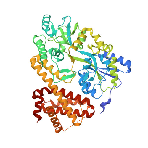
    Characterizing protein-ligand interactions is crucial to understanding cellular metabolism and guiding drug discovery and development. Herein, we explore complementing hydrogen/deuterium exchange mass spectrometry (HDX-MS) with a recently developed Fenton chemistry-based approach to protein oxidative footprinting mass spectrometry (OX-MS) to discriminate the binding of small-molecule therapeutics. Using drug-dependent perturbation as the experimental report, this combination of techniques more clearly differentiates the in-solution binding profiles of Venetoclax (ABT-199, GDC-0199-AbbVie and Genentech) and a drug candidate S55746 (Servier) to the apoptotic regulatory protein Bcl-2 than either technique alone. These results highlight the value of combining these methods to compare compounds in drug discovery and development. To better understand the structural context of the HDX-MS and OX-MS drug-dependent perturbations, we mapped these data on Bcl-2-Venetoclax and Bcl-2-S55746 cocrystal structures and compared these results with the structure of apo Bcl-2. HDX-MS shows that Venetoclax more strongly impacts the protein backbone compared to S55746. OX-MS reveals oxidation perturbations rationalized by direct side-chain protection as well as by crystallographically observed drug-induced protein restructuring. Both methods report the perturbation of some, but not all, residues mapped within 4 Å of the bound drugs in the crystal structures. Concordant characterization of backbone and side-chain accessibility will enhance our understanding of in-solution protein structure dynamics and protein-ligand interactions during drug discovery, development, and characterization, particularly when high-resolution structures are lacking.


  • Organizational Affiliation
    • Department of Biochemistry, Albert Einstein College of Medicine, Bronx, New York 10461, United States.

Macromolecule Content 

  • Total Structure Weight: 61.26 kDa 
  • Atom Count: 4,096 
  • Modeled Residue Count: 503 
  • Deposited Residue Count: 547 
  • Unique protein chains: 1

Macromolecules

Find similar proteins by:|  3D Structure
Entity ID: 1
MoleculeChains  Sequence LengthOrganismDetailsImage
Maltose/maltodextrin-binding periplasmic protein fused to apoptosis regulator Bcl-2/Bcl-xL chimera547Escherichia coli K-12Homo sapiensMutation(s): 5 
Gene Names: malEb4034JW3994BCL2BCL2L1BCL2LBCLX
UniProt & NIH Common Fund Data Resources
Find proteins for P0AEX9 (Escherichia coli (strain K12))
Explore P0AEX9 
Go to UniProtKB:  P0AEX9
Find proteins for P10415 (Homo sapiens)
Explore P10415 
Go to UniProtKB:  P10415
PHAROS:  P10415
GTEx:  ENSG00000171791 
Find proteins for Q07817 (Homo sapiens)
Explore Q07817 
Go to UniProtKB:  Q07817
PHAROS:  Q07817
GTEx:  ENSG00000171552 
Entity Groups
Sequence Clusters30% Identity50% Identity70% Identity90% Identity95% Identity100% Identity
UniProt GroupsQ07817P10415P0AEX9
Sequence Annotations
Expand
Reference Sequence

Experimental Data & Validation

Experimental Data

  • Method: X-RAY DIFFRACTION
  • Resolution: 2.09 Å
  • R-Value Free:  0.264 (Depositor), 0.263 (DCC) 
  • R-Value Work:  0.204 (Depositor), 0.204 (DCC) 
  • R-Value Observed: 0.206 (Depositor) 
Space Group: P 21 21 21
Unit Cell:
Length ( Å )Angle ( ˚ )
a = 37.07α = 90
b = 88.95β = 90
c = 140.94γ = 90
Software Package:
Software NamePurpose
PHENIXrefinement
autoPROCdata reduction
PHASERphasing
autoPROCdata scaling

Structure Validation

View Full Validation Report


Entry History 

& Funding Information

Deposition Data


Funding OrganizationLocationGrant Number
Not funded--

Revision History  (Full details and data files)

  • Version 1.0: 2024-10-02
    Type: Initial release
  • Version 1.1: 2025-03-05
    Changes: Database references, Structure summary
  • Version 1.2: 2025-03-12
    Changes: Database references