8VUW | pdb_00008vuw

ELIC5 with cysteamine in 2:1:1 POPC:POPE:POPG nanodisc in open conformation


Experimental Data Snapshot

  • Method: ELECTRON MICROSCOPY
  • Resolution: 3.19 Å
  • Aggregation State: PARTICLE 
  • Reconstruction Method: SINGLE PARTICLE 

wwPDB Validation 3D Report Full Report

Validation slider image for 8VUW

This is version 1.0 of the entry. See complete history

Literature

Open-channel structure of a pentameric ligand-gated ion channel reveals a mechanism of leaflet-specific phospholipid modulation.

Petroff, J.T.Dietzen, N.M.Santiago-McRae, E.Deng, B.Washington, M.S.Chen, L.J.Trent Moreland, K.Deng, Z.Rau, M.Fitzpatrick, J.A.J.Yuan, P.Joseph, T.T.Henin, J.Brannigan, G.Cheng, W.W.L.

(2022) Nat Commun 13: 7017

  • DOI: https://doi.org/10.1038/s41467-022-34813-5
  • Primary Citation Related Structures: 
    8D63, 8D64, 8D65, 8D66, 8D67, 8VUW

  • PubMed Abstract: 

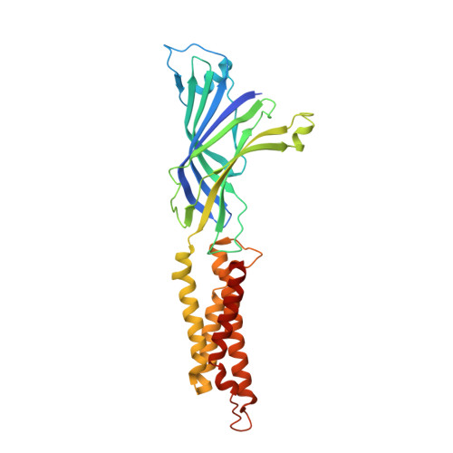
    Pentameric ligand-gated ion channels (pLGICs) mediate synaptic transmission and are sensitive to their lipid environment. The mechanism of phospholipid modulation of any pLGIC is not well understood. We demonstrate that the model pLGIC, ELIC (Erwinia ligand-gated ion channel), is positively modulated by the anionic phospholipid, phosphatidylglycerol, from the outer leaflet of the membrane. To explore the mechanism of phosphatidylglycerol modulation, we determine a structure of ELIC in an open-channel conformation. The structure shows a bound phospholipid in an outer leaflet site, and structural changes in the phospholipid binding site unique to the open-channel. In combination with streamlined alchemical free energy perturbation calculations and functional measurements in asymmetric liposomes, the data support a mechanism by which an anionic phospholipid stabilizes the activated, open-channel state of a pLGIC by specific, state-dependent binding to this site.


  • Organizational Affiliation
    • Department of Anesthesiology, Washington University School of Medicine, Saint Louis, MO, USA.

Macromolecule Content 

  • Total Structure Weight: 189.19 kDa 
  • Atom Count: 13,055 
  • Modeled Residue Count: 1,555 
  • Deposited Residue Count: 1,610 
  • Unique protein chains: 1

Macromolecules

Find similar proteins by:|  3D Structure
Entity ID: 1
MoleculeChains  Sequence LengthOrganismDetailsImage
Erwinia chrysanthemi ligand-gated ion channel
A, B, C, D, E
322Dickeya chrysanthemiMutation(s): 5 
Membrane Entity: Yes 
UniProt
Find proteins for P0C7B7 (Dickeya chrysanthemi)
Explore P0C7B7 
Go to UniProtKB:  P0C7B7
Entity Groups
Sequence Clusters30% Identity50% Identity70% Identity90% Identity95% Identity100% Identity
UniProt GroupP0C7B7
Sequence Annotations
Expand
Reference Sequence

Small Molecules

Ligands 2 Unique
IDChains Name / Formula / InChI Key2D Diagram3D Interactions
PGW
(Subject of Investigation/LOI)

Query on PGW


Download:Ideal Coordinates CCD File
F [auth A],
H [auth A],
I [auth B],
K [auth C],
M [auth D]
(1R)-2-{[(S)-{[(2S)-2,3-dihydroxypropyl]oxy}(hydroxy)phosphoryl]oxy}-1-[(hexadecanoyloxy)methyl]ethyl (9Z)-octadec-9-enoate
C40 H77 O10 P
PAZGBAOHGQRCBP-HGWHEPCSSA-N
DHL
(Subject of Investigation/LOI)

Query on DHL


Download:Ideal Coordinates CCD File
G [auth A],
J [auth B],
L [auth C],
N [auth D],
O [auth E]
2-AMINO-ETHANETHIOL
C2 H7 N S
UFULAYFCSOUIOV-UHFFFAOYSA-N

Experimental Data & Validation

Experimental Data

  • Method: ELECTRON MICROSCOPY
  • Resolution: 3.19 Å
  • Aggregation State: PARTICLE 
  • Reconstruction Method: SINGLE PARTICLE 

Structure Validation

View Full Validation Report


Entry History 

& Funding Information

Deposition Data


Funding OrganizationLocationGrant Number
National Institutes of Health/National Institute of General Medical Sciences (NIH/NIGMS)United StatesGM137957

Revision History  (Full details and data files)

  • Version 1.0: 2024-02-07
    Type: Initial release