8VRH | pdb_00008vrh

E. coli peptidyl-prolyl cis-trans isomerase containing delta2-monofluoro-leucines


Experimental Data Snapshot

  • Method: X-RAY DIFFRACTION
  • Resolution: 1.80 Å
  • R-Value Free: 
    0.247 (Depositor), 0.262 (DCC) 
  • R-Value Work: 
    0.208 (Depositor), 0.223 (DCC) 
  • R-Value Observed: 
    0.209 (Depositor) 

Starting Model: experimental
View more details

wwPDB Validation 3D Report Full Report

Validation slider image for 8VRH

This is version 1.2 of the entry. See complete history

Literature

Conformational Preferences of the Non-Canonical Amino Acids (2 S ,4 S )-5-Fluoroleucine, (2 S ,4 R )-5-Fluoroleucine, and 5,5'-Difluoroleucine in a Protein.

Frkic, R.L.Tan, Y.J.Abdelkader, E.H.Maleckis, A.Tarcoveanu, E.Nitsche, C.Otting, G.Jackson, C.J.

(2024) Biochemistry 63: 1388-1394

  • DOI: https://doi.org/10.1021/acs.biochem.4c00081
  • Primary Citation Related Structures: 
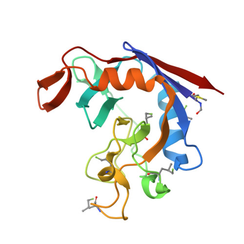
    8VRG, 8VRH, 8VRI

  • PubMed Abstract: 

    Proteins produced with leucine analogues, where CH 2 F groups substitute specific methyl groups, can readily be probed by 19 F NMR spectroscopy. As CF and CH groups are similar in hydrophobicity and size, fluorinated leucines are expected to cause minimal structural perturbation, but the impact of fluorine on the rotational freedom of CH 2 F groups is unclear. We present high-resolution crystal structures of Escherichia coli peptidyl-prolyl cis - trans isomerase B (PpiB) prepared with uniform high-level substitution of leucine by (2 S ,4 S )-5-fluoroleucine, (2 S ,4 R )-5-fluoroleucine, or 5,5'-difluoroleucine. Apart from the fluorinated leucine residues, the structures show complete structural conservation of the protein backbone and the amino acid side chains except for a single isoleucine side chain located next to a fluorine atom in the hydrophobic core of the protein. The carbon skeletons of the fluorinated leucine side chains are also mostly conserved. The CH 2 F groups show a strong preference for staggered rotamers and often appear locked into single rotamers. Substitution of leucine CH 3 groups for CH 2 F groups is thus readily tolerated in the three-dimensional (3D) structure of a protein, and the rotation of CH 2 F groups can be halted at cryogenic temperatures.


  • Organizational Affiliation
    • ARC Centre of Excellence for Innovations in Peptide & Protein Science, Research School of Chemistry, Australian National University, Canberra, ACT 2601, Australia.

Macromolecule Content 

  • Total Structure Weight: 74.71 kDa 
  • Atom Count: 5,638 
  • Modeled Residue Count: 655 
  • Deposited Residue Count: 656 
  • Unique protein chains: 1

Macromolecules

Find similar proteins by:|  3D Structure
Entity ID: 1
MoleculeChains  Sequence LengthOrganismDetailsImage
Peptidyl-prolyl cis-trans isomerase B
A, B, C, D
164Escherichia coliMutation(s): 0 
Gene Names: ppiBb0525JW0514
EC: 5.2.1.8
UniProt
Find proteins for P23869 (Escherichia coli (strain K12))
Explore P23869 
Go to UniProtKB:  P23869
Entity Groups
Sequence Clusters30% Identity50% Identity70% Identity90% Identity95% Identity100% Identity
UniProt GroupP23869
Sequence Annotations
Expand
Reference Sequence

Small Molecules

Ligands 5 Unique
IDChains Name / Formula / InChI Key2D Diagram3D Interactions
XPE

Query on XPE


Download:Ideal Coordinates CCD File
M [auth C]3,6,9,12,15,18,21,24,27-NONAOXANONACOSANE-1,29-DIOL
C20 H42 O11
DTPCFIHYWYONMD-UHFFFAOYSA-N
1PE

Query on 1PE


Download:Ideal Coordinates CCD File
L [auth B]PENTAETHYLENE GLYCOL
C10 H22 O6
JLFNLZLINWHATN-UHFFFAOYSA-N
PGE

Query on PGE


Download:Ideal Coordinates CCD File
H [auth A],
O [auth D]
TRIETHYLENE GLYCOL
C6 H14 O4
ZIBGPFATKBEMQZ-UHFFFAOYSA-N
PEG

Query on PEG


Download:Ideal Coordinates CCD File
J [auth B]DI(HYDROXYETHYL)ETHER
C4 H10 O3
MTHSVFCYNBDYFN-UHFFFAOYSA-N
EDO

Query on EDO


Download:Ideal Coordinates CCD File
E [auth A]
F [auth A]
G [auth A]
I [auth A]
K [auth B]
E [auth A],
F [auth A],
G [auth A],
I [auth A],
K [auth B],
N [auth C],
P [auth D]
1,2-ETHANEDIOL
C2 H6 O2
LYCAIKOWRPUZTN-UHFFFAOYSA-N
Modified Residues  2 Unique
IDChains TypeFormula2D DiagramParent
A1ADO
Query on A1ADO
A, B, C, D
L-PEPTIDE LINKINGC6 H12 F N O2LEU
FME
Query on FME
A, B, C, D
L-PEPTIDE LINKINGC6 H11 N O3 SMET

Experimental Data & Validation

Experimental Data

  • Method: X-RAY DIFFRACTION
  • Resolution: 1.80 Å
  • R-Value Free:  0.247 (Depositor), 0.262 (DCC) 
  • R-Value Work:  0.208 (Depositor), 0.223 (DCC) 
  • R-Value Observed: 0.209 (Depositor) 
Space Group: P 21 21 21
Unit Cell:
Length ( Å )Angle ( ˚ )
a = 79.022α = 90
b = 83.003β = 90
c = 121.642γ = 90
Software Package:
Software NamePurpose
PHENIXrefinement
Aimlessdata scaling
CrysalisProdata reduction
PHASERphasing

Structure Validation

View Full Validation Report


Entry History 

& Funding Information

Deposition Data


Funding OrganizationLocationGrant Number
Australian Research Council (ARC)AustraliaCE200100012

Revision History  (Full details and data files)

  • Version 1.0: 2024-05-22
    Type: Initial release
  • Version 1.1: 2024-05-29
    Changes: Database references, Derived calculations
  • Version 1.2: 2024-06-19
    Changes: Database references