8VMK | pdb_00008vmk

A crystal structure of heme-dependent tyrosine hydroxylase complexed with a substrate analog, 3-(4-hydroxyphenyl)propionic acid


Experimental Data Snapshot

  • Method: X-RAY DIFFRACTION
  • Resolution: 1.56 Å
  • R-Value Free: 
    0.213 (Depositor), 0.213 (DCC) 
  • R-Value Work: 
    0.187 (Depositor), 0.187 (DCC) 
  • R-Value Observed: 
    0.188 (Depositor) 

Starting Model: experimental
View more details

wwPDB Validation 3D Report Full Report

Validation slider image for 8VMK

Ligand Structure Quality Assessment 


This is version 1.1 of the entry. See complete history

Literature

Substrate Analogs Implicate a Free Radical Pathway in Tyrosine Hydroxylase Catalysis.

Traore, E.S.Wang, Y.Griffith, W.P.Liu, A.

(2025) ACS Catal 15: 18270-18281

  • DOI: https://doi.org/10.1021/acscatal.5c05776
  • Primary Citation Related Structures: 
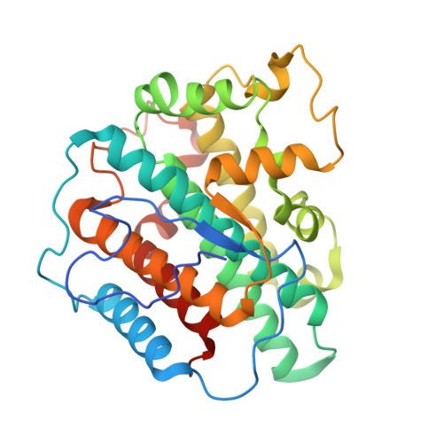
    8VMK

  • PubMed Abstract: 

    Heme-dependent tyrosine hydroxylases (TyrH) are critical enzymes in catecholamine biosynthesis, yet the role of the substrate's α -amino group in their monooxygenation mechanism has been unclear. Using 3-(4-hydroxyphenyl)propionic acid (HPPA), an l-tyrosine analog lacking the α -amino group, we observed a distinct dimerization pathway that competes with the expected hydroxylation reaction. Several lines of evidence confirm that this process originates from a radical intermediate. First, the formation of this (HPPA) 2 dimer is selectively inhibited by a free radical scavenger. Second, 18 O-labeling experiments show phenolic oxygen scrambling, indicating a disruption of substrate aromaticity during catalysis. Finally, EPR spectroscopy using nitrosobenzene as a substrate analog revealed a substrate-based free radical. This mechanistic divergence clarifies the role of the α -amino group. Its absence in HPPA creates a kinetic bottleneck for the final O atom transfer step, allowing a fraction of the substrate radical to form the off-pathway dimer. Thus, the native substrate's α -amino group acts as a crucial kinetic modulator, ensuring the rapid and efficient commitment of the substrate radical to productive hydroxylation. These results collectively establish a peroxidase-like free radical pathway for TyrH and reveal the nonessential yet significant role the amino group plays in controlling reaction outcomes.


  • Organizational Affiliation
    • Department of Chemistry, University of Texas at San Antonio, San Antonio, Texas 78249, United States.

Macromolecule Content 

  • Total Structure Weight: 71.06 kDa 
  • Atom Count: 5,417 
  • Modeled Residue Count: 604 
  • Deposited Residue Count: 632 
  • Unique protein chains: 1

Macromolecules

Find similar proteins by:|  3D Structure
Entity ID: 1
MoleculeChains  Sequence LengthOrganismDetailsImage
Heme-dependent L-tyrosine hydroxylase
A, B
316Streptomyces sclerotialusMutation(s): 0 
UniProt
Find proteins for A0ACD6BA76 (Streptomyces sp. ICBB 8177)
Explore A0ACD6BA76 
Go to UniProtKB:  A0ACD6BA76
Entity Groups
Sequence Clusters30% Identity50% Identity70% Identity90% Identity95% Identity100% Identity
UniProt GroupA0ACD6BA76
Sequence Annotations
Expand
Reference Sequence

Small Molecules

Ligands 3 Unique
IDChains Name / Formula / InChI Key2D Diagram3D Interactions
HEM
(Subject of Investigation/LOI)

Query on HEM


Download:Ideal Coordinates CCD File
D [auth A],
G [auth B]
PROTOPORPHYRIN IX CONTAINING FE
C34 H32 Fe N4 O4
KABFMIBPWCXCRK-RGGAHWMASA-L
HPP
(Subject of Investigation/LOI)

Query on HPP


Download:Ideal Coordinates CCD File
C [auth A],
F [auth B]
HYDROXYPHENYL PROPIONIC ACID
C9 H10 O3
NMHMNPHRMNGLLB-UHFFFAOYSA-N
TRS

Query on TRS


Download:Ideal Coordinates CCD File
E [auth A],
H [auth B]
2-AMINO-2-HYDROXYMETHYL-PROPANE-1,3-DIOL
C4 H12 N O3
LENZDBCJOHFCAS-UHFFFAOYSA-O

Experimental Data & Validation

Experimental Data

  • Method: X-RAY DIFFRACTION
  • Resolution: 1.56 Å
  • R-Value Free:  0.213 (Depositor), 0.213 (DCC) 
  • R-Value Work:  0.187 (Depositor), 0.187 (DCC) 
  • R-Value Observed: 0.188 (Depositor) 
Space Group: P 1 21 1
Unit Cell:
Length ( Å )Angle ( ˚ )
a = 47.634α = 90
b = 129.819β = 93.86
c = 48.42γ = 90
Software Package:
Software NamePurpose
PHENIXrefinement
HKL-2000data scaling
HKL-2000data reduction
PHASERphasing

Structure Validation

View Full Validation Report


Ligand Structure Quality Assessment 


Entry History 

& Funding Information

Deposition Data


Funding OrganizationLocationGrant Number
National Institutes of Health/National Institute of General Medical Sciences (NIH/NIGMS)United StatesR01GM108988

Revision History  (Full details and data files)

  • Version 1.0: 2025-08-27
    Type: Initial release
  • Version 1.1: 2026-03-25
    Changes: Database references