8VMB | pdb_00008vmb

The crystal structure of rhinovirus C15 RNA replication element sB-loop mutant in complex with Fab BL3-6


Experimental Data Snapshot

  • Method: X-RAY DIFFRACTION
  • Resolution: 2.97 Å
  • R-Value Free: 
    0.251 (Depositor), 0.253 (DCC) 
  • R-Value Work: 
    0.210 (Depositor), 0.213 (DCC) 
  • R-Value Observed: 
    0.214 (Depositor) 

Starting Model: experimental
View more details

wwPDB Validation 3D Report Full Report

Validation slider image for 8VMB

This is version 1.3 of the entry. See complete history

Literature

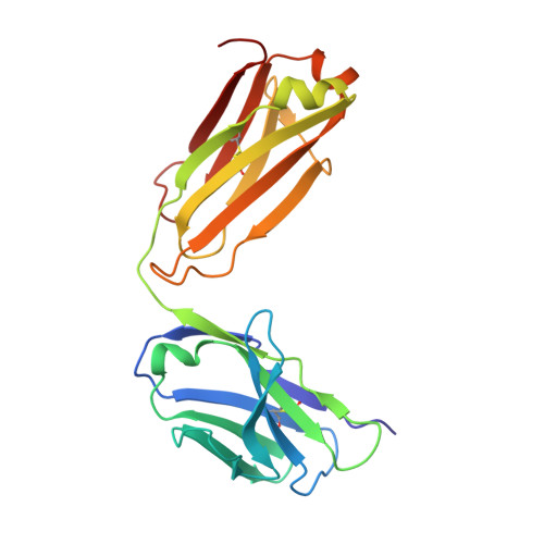
Structural basis for a highly conserved RNA-mediated enteroviral genome replication.

Das, N.K.Vogt, J.Patel, A.Banna, H.A.Koirala, D.

(2024) Nucleic Acids Res 52: 11218-11233

  • DOI: https://doi.org/10.1093/nar/gkae627
  • Primary Citation Related Structures: 
    8VM8, 8VM9, 8VMA, 8VMB

  • PubMed Abstract: 

    Enteroviruses contain conserved RNA structures at the extreme 5' end of their genomes that recruit essential proteins 3CD and PCBP2 to promote genome replication. However, the high-resolution structures and mechanisms of these replication-linked RNAs (REPLRs) are limited. Here, we determined the crystal structures of the coxsackievirus B3 and rhinoviruses B14 and C15 REPLRs at 1.54, 2.2 and 2.54 Å resolution, revealing a highly conserved H-type four-way junction fold with co-axially stacked sA-sD and sB-sC helices that are stabilized by a long-range A•C•U base-triple. Such conserved features observed in the crystal structures also allowed us to predict the models of several other enteroviral REPLRs using homology modeling, which generated models almost identical to the experimentally determined structures. Moreover, our structure-guided binding studies with recombinantly purified full-length human PCBP2 showed that two previously proposed binding sites, the sB-loop and 3' spacer, reside proximally and bind a single PCBP2. Additionally, the DNA oligos complementary to the 3' spacer, the high-affinity PCBP2 binding site, abrogated its interactions with enteroviral REPLRs, suggesting the critical roles of this single-stranded region in recruiting PCBP2 for enteroviral genome replication and illuminating the promising prospects of developing therapeutics against enteroviral infections targeting this replication platform.


  • Organizational Affiliation
    • Department of Chemistry and Biochemistry, University of Maryland, Baltimore County, Baltimore, MD 21250, USA.

Macromolecule Content 

  • Total Structure Weight: 75.37 kDa 
  • Atom Count: 5,105 
  • Modeled Residue Count: 526 
  • Deposited Residue Count: 533 
  • Unique protein chains: 2
  • Unique nucleic acid chains: 1

Macromolecules


Find similar proteins by:|  3D Structure
Entity ID: 2
MoleculeChains  Sequence LengthOrganismDetailsImage
Heavy Chain of Fab BL3-6B [auth H]233Homo sapiensMutation(s): 0 
Find similar proteins by:|  3D Structure
Entity ID: 3
MoleculeChains  Sequence LengthOrganismDetailsImage
Light Chain of Fab BL3-6C [auth L]215Homo sapiensMutation(s): 0 
Find similar nucleic acids by:  (by identity cutoff) 
Entity ID: 1
MoleculeChains LengthOrganismImage
RNA (83-MER)A [auth R]85synthetic construct
Sequence Annotations
Expand
Reference Sequence

Experimental Data & Validation

Experimental Data

  • Method: X-RAY DIFFRACTION
  • Resolution: 2.97 Å
  • R-Value Free:  0.251 (Depositor), 0.253 (DCC) 
  • R-Value Work:  0.210 (Depositor), 0.213 (DCC) 
  • R-Value Observed: 0.214 (Depositor) 
Space Group: I 21 21 21
Unit Cell:
Length ( Å )Angle ( ˚ )
a = 103.216α = 90
b = 121.847β = 90
c = 174.664γ = 90
Software Package:
Software NamePurpose
PHENIXphasing
PHENIXrefinement
RAPDdata reduction
RAPDdata processing
RAPDdata scaling

Structure Validation

View Full Validation Report


Entry History 

& Funding Information

Deposition Data


Funding OrganizationLocationGrant Number
National Science Foundation (NSF, United States)United StatesMCB-2236996

Revision History  (Full details and data files)

  • Version 1.0: 2024-07-24
    Type: Initial release
  • Version 1.1: 2024-08-07
    Changes: Database references
  • Version 1.2: 2024-10-23
    Changes: Structure summary
  • Version 1.3: 2024-10-30
    Changes: Database references