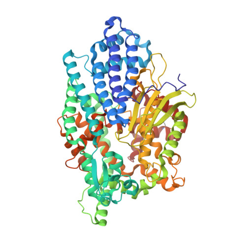
8VJY

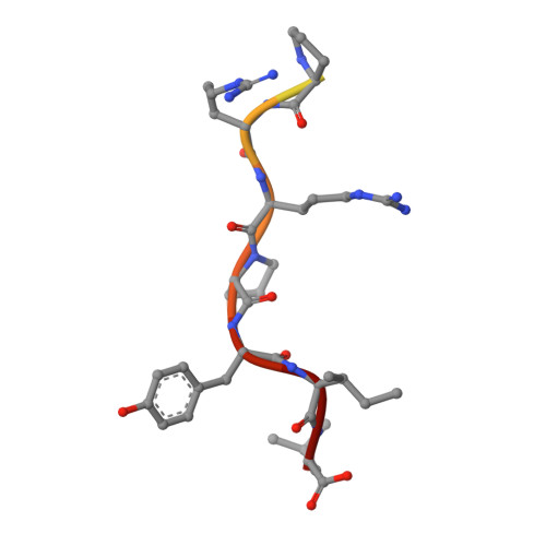
Structure of Human Neurolysin in complex with Neurotensin peptide

  • Classification: HYDROLASE
  • Organism(s): Homo sapiens
  • Expression System: Escherichia coli
  • Mutation(s): No 

  • Deposited: 2024-01-08 Released: 2024-08-21 
  • Deposition Author(s): Shi, K., Aihara, H.
  • Funding Organization(s): National Institutes of Health/National Institute of Neurological Disorders and Stroke (NIH/NINDS), National Institutes of Health/National Institute of General Medical Sciences (NIH/NIGMS)

Experimental Data Snapshot

  • Method: X-RAY DIFFRACTION
  • Resolution: 1.95 Å
  • R-Value Free: 0.234 
  • R-Value Work: 0.198 
  • R-Value Observed: 0.199 

Starting Model: experimental
View more details

wwPDB Validation   3D Report Full Report


This is version 1.0 of the entry. See complete history


Literature

Structural basis of divergent substrate recognition and inhibition of human neurolysin.

Shi, K.Bagchi, S.Bickel, J.Esfahani, S.H.Yin, L.Cheng, T.Karamyan, V.T.Aihara, H.

(2024) Sci Rep 14: 18420-18420

  • DOI: https://doi.org/10.1038/s41598-024-67639-w
  • Primary Citation of Related Structures:  
    8VJU, 8VJV, 8VJX, 8VJY

  • PubMed Abstract: 

    A zinc metallopeptidase neurolysin (Nln) processes diverse bioactive peptides to regulate signaling in the mammalian nervous system. To understand how Nln interacts with various peptides with dissimilar sequences, we determined crystal structures of Nln in complex with diverse peptides including dynorphins, angiotensin, neurotensin, and bradykinin. The structures show that Nln binds these peptides in a large dumbbell-shaped interior cavity constricted at the active site, making minimal structural changes to accommodate different peptide sequences. The structures also show that Nln readily binds similar peptides with distinct registers, which can determine whether the peptide serves as a substrate or a competitive inhibitor. We analyzed the activities and binding of Nln toward various forms of dynorphin A peptides, which highlights the promiscuous nature of peptide binding and shows how dynorphin A (1-13) potently inhibits the Nln activity while dynorphin A (1-8) is efficiently cleaved. Our work provides insights into the broad substrate specificity of Nln and may aid in the future design of small molecule modulators for Nln.


  • Organizational Affiliation

    Department of Biochemistry, Molecular Biology and Biophysics, University of Minnesota, Minneapolis, MN, 55455, USA.


Macromolecules
Find similar proteins by:  (by identity cutoff)  |  3D Structure
Entity ID: 1
MoleculeChains Sequence LengthOrganismDetailsImage
Neurolysin, mitochondrial
A, C
667Homo sapiensMutation(s): 0 
Gene Names: NLNAGTBPKIAA1226
EC: 3.4.24.16
UniProt & NIH Common Fund Data Resources
Find proteins for Q9BYT8 (Homo sapiens)
Explore Q9BYT8 
Go to UniProtKB:  Q9BYT8
PHAROS:  Q9BYT8
GTEx:  ENSG00000123213 
Entity Groups  
Sequence Clusters30% Identity50% Identity70% Identity90% Identity95% Identity100% Identity
UniProt GroupQ9BYT8
Sequence Annotations
Expand
  • Reference Sequence

Find similar proteins by:  Sequence   |   3D Structure  

Entity ID: 2
MoleculeChains Sequence LengthOrganismDetailsImage
Neurotensin
B, D
14Homo sapiensMutation(s): 0 
Entity Groups  
Sequence Clusters30% Identity50% Identity70% Identity90% Identity95% Identity100% Identity
Sequence Annotations
Expand
  • Reference Sequence
Small Molecules
Ligands 2 Unique
IDChains Name / Formula / InChI Key2D Diagram3D Interactions
ZN
Query on ZN

Download Ideal Coordinates CCD File 
E [auth A],
R [auth C]
ZINC ION
Zn
PTFCDOFLOPIGGS-UHFFFAOYSA-N
EDO
Query on EDO

Download Ideal Coordinates CCD File 
AA [auth C]
BA [auth C]
F [auth A]
G [auth A]
H [auth A]
AA [auth C],
BA [auth C],
F [auth A],
G [auth A],
H [auth A],
I [auth A],
J [auth A],
K [auth A],
L [auth A],
M [auth A],
N [auth A],
O [auth A],
P [auth A],
Q [auth A],
S [auth C],
T [auth C],
U [auth C],
V [auth C],
W [auth C],
X [auth C],
Y [auth C],
Z [auth C]
1,2-ETHANEDIOL
C2 H6 O2
LYCAIKOWRPUZTN-UHFFFAOYSA-N
Modified Residues  1 Unique
IDChains TypeFormula2D DiagramParent
CME
Query on CME
A, C
L-PEPTIDE LINKINGC5 H11 N O3 S2CYS
Experimental Data & Validation

Experimental Data

  • Method: X-RAY DIFFRACTION
  • Resolution: 1.95 Å
  • R-Value Free: 0.234 
  • R-Value Work: 0.198 
  • R-Value Observed: 0.199 
  • Space Group: P 21 21 21
Unit Cell:
Length ( Å )Angle ( ˚ )
a = 59.712α = 90
b = 150.576β = 90
c = 188.799γ = 90
Software Package:
Software NamePurpose
PHENIXrefinement
STARANISOdata scaling
XDSdata reduction
PHASERphasing

Structure Validation

View Full Validation Report


Entry History & Funding Information

Deposition Data


Funding OrganizationLocationGrant Number
National Institutes of Health/National Institute of Neurological Disorders and Stroke (NIH/NINDS)United StatesR01NS106879
National Institutes of Health/National Institute of General Medical Sciences (NIH/NIGMS)United StatesR35GM118047

Revision History  (Full details and data files)

  • Version 1.0: 2024-08-21
    Type: Initial release