Structural insights into the convergent evolution of sulfoxide synthase EgtB-IV, an ergothioneine-biosynthetic homolog of ovothiol synthase OvoA.
Ireland, K.A., Kayrouz, C.M., Abbott, M.L., Seyedsayamdost, M.R., Davis, K.M.(2024) Structure 32: 2013-2022.e5
- PubMed: 39216472 Search on PubMed
- DOI: https://doi.org/10.1016/j.str.2024.08.006
- Primary Citation Related Structures:
8VIG, 8VIH, 8VII, 8VIK, 8VIL - PubMed Abstract:
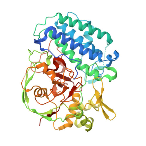
Non-heme iron-dependent sulfoxide/selenoxide synthases (NHISS) constitute a unique metalloenzyme class capable of installing a C-S/Se bond onto histidine to generate thio/selenoimidazole antioxidants, such as ergothioneine and ovothiol. These natural products are increasingly recognized for their health benefits. Among associated ergothioneine-biosynthetic enzymes, type IV EgtBs stand out, as they exhibit low sequence similarity with other EgtB subfamilies due to their recent divergence from the ovothiol-biosynthetic enzyme OvoA. Herein, we present crystal structures of two representative EgtB-IV enzymes, offering insights into the basis for this evolutionary convergence and enhancing our understanding of NHISS active site organization more broadly. The ability to interpret how key residues modulate substrate specificity and regioselectivity has implications for downstream identification of divergent reactivity within the NHISS family. To this end, we identify a previously unclassified clade of OvoA-like enzymes with a seemingly hybrid set of characteristics, suggesting they may represent an evolutionary intermediate between OvoA and EgtB-IV.
- Department of Chemistry, Emory University, Atlanta, GA 30322, USA.
Organizational Affiliation: