8VE8 | pdb_00008ve8

Lineage IV Lassa virus glycoprotein (Josiah) in complex with rabbit polyclonal antibody (GP1-A epitope)


Experimental Data Snapshot

  • Method: ELECTRON MICROSCOPY
  • Resolution: 2.80 Å
  • Aggregation State: PARTICLE 
  • Reconstruction Method: SINGLE PARTICLE 

wwPDB Validation 3D Report Full Report

Validation slider image for 8VE8

This is version 1.2 of the entry. See complete history

Literature

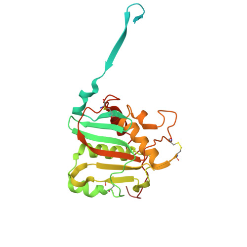
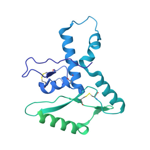
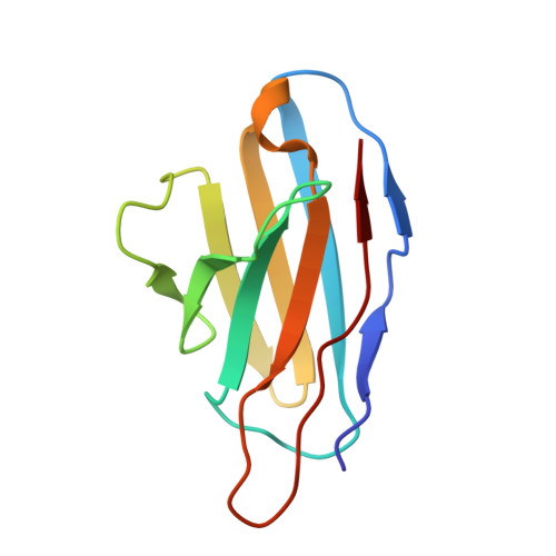
Defining bottlenecks and opportunities for Lassa virus neutralization by structural profiling of vaccine-induced polyclonal antibody responses.

Brouwer, P.J.M.Perrett, H.R.Beaumont, T.Nijhuis, H.Kruijer, S.Burger, J.A.Bontjer, I.Lee, W.H.Ferguson, J.A.Schauflinger, M.Muller-Krauter, H.Sanders, R.W.Strecker, T.van Gils, M.J.Ward, A.B.

(2024) Cell Rep 43: 114708-114708

  • DOI: https://doi.org/10.1016/j.celrep.2024.114708
  • Primary Citation Related Structures: 
    8TYC, 8TYE, 8VCV, 8VE8, 9CJ7, 9CJ8, 9CK7, 9CK8

  • PubMed Abstract: 

    Lassa fever continues to be a major public health burden in West Africa, yet effective therapies or vaccines are lacking. The isolation of protective neutralizing antibodies against the Lassa virus glycoprotein complex (GPC) justifies the development of vaccines that can elicit strong neutralizing antibody responses. However, Lassa vaccine candidates have generally been unsuccessful at doing so, and the associated antibody responses to these vaccines remain poorly characterized. Here, we establish an electron microscopy-based epitope mapping workflow that enables high-resolution structural characterization of polyclonal antibodies to the GPC. By applying this method to rabbits vaccinated with a recombinant GPC vaccine and a GPC-derived virus-like particle, we reveal determinants of neutralization that involve epitopes of the GPC-A competition cluster. Furthermore, by identifying undescribed immunogenic off-target epitopes, we expose the challenges that recombinant GPC vaccines face. By enabling detailed polyclonal antibody characterization, our work ushers in a next generation of more rational Lassa vaccine design.


  • Organizational Affiliation
    • Department of Integrative Structural and Computational Biology, Scripps Research, La Jolla, CA 92037, USA.

Macromolecule Content 

  • Total Structure Weight: 254.39 kDa 
  • Atom Count: 10,327 
  • Modeled Residue Count: 1,260 
  • Deposited Residue Count: 2,220 
  • Unique protein chains: 4

Macromolecules

Find similar proteins by:|  3D Structure
Entity ID: 1
MoleculeChains  Sequence LengthOrganismDetailsImage
Glycoprotein G1
A, B, C
259Lassa virus JosiahMutation(s): 0 
Gene Names: GPC
UniProt
Find proteins for P08669 (Lassa virus (strain Mouse/Sierra Leone/Josiah/1976))
Explore P08669 
Go to UniProtKB:  P08669
Entity Groups
Sequence Clusters30% Identity50% Identity70% Identity90% Identity95% Identity100% Identity
UniProt GroupP08669
Glycosylation
Glycosylation Sites: 7
Sequence Annotations
Expand
Reference Sequence
Find similar proteins by:|  3D Structure
Entity ID: 2
MoleculeChains  Sequence LengthOrganismDetailsImage
Glycoprotein G2D [auth a],
E [auth b],
F [auth c]
406Lassa virus JosiahMutation(s): 0 
Gene Names: GPCGP-C
UniProt
Find proteins for P08669 (Lassa virus (strain Mouse/Sierra Leone/Josiah/1976))
Explore P08669 
Go to UniProtKB:  P08669
Entity Groups
Sequence Clusters30% Identity50% Identity70% Identity90% Identity95% Identity100% Identity
UniProt GroupP08669
Glycosylation
Glycosylation Sites: 4
Sequence Annotations
Expand
Reference Sequence
Find similar proteins by:|  3D Structure
Entity ID: 3
MoleculeChains  Sequence LengthOrganismDetailsImage
Rabbit polyclonal Fv light chainG [auth L]106Oryctolagus cuniculusMutation(s): 0 
Find similar proteins by:|  3D Structure
Entity ID: 4
MoleculeChains  Sequence LengthOrganismDetailsImage
Rabbit polyclonal Fv heavy chain119Oryctolagus cuniculusMutation(s): 0 

Oligosaccharides

Help  
Entity ID: 5
MoleculeChains Length2D Diagram GlycosylationD Interactions
2-acetamido-2-deoxy-beta-D-glucopyranose-(1-4)-2-acetamido-2-deoxy-beta-D-glucopyranose
I [auth D],
J [auth E],
L [auth G],
M [auth I],
O [auth K],
I [auth D],
J [auth E],
L [auth G],
M [auth I],
O [auth K],
P [auth M],
S [auth P],
T [auth Q],
U [auth R],
X [auth U]
2N-Glycosylation
Glycosylation Resources
GlyTouCan: G42666HT
GlyCosmos: G42666HT
GlyGen: G42666HT
Entity ID: 6
MoleculeChains Length2D Diagram GlycosylationD Interactions
2-acetamido-2-deoxy-beta-D-glucopyranose-(1-4)-[alpha-L-fucopyranose-(1-6)]2-acetamido-2-deoxy-beta-D-glucopyranoseAA [auth X],
K [auth F],
N [auth J],
R [auth O],
V [auth S]
3N-Glycosylation
Glycosylation Resources
GlyTouCan: G21290RB
GlyCosmos: G21290RB
GlyGen: G21290RB
Entity ID: 7
MoleculeChains Length2D Diagram GlycosylationD Interactions
alpha-D-mannopyranose-(1-2)-alpha-D-mannopyranose-(1-3)-[alpha-D-mannopyranose-(1-6)]beta-D-mannopyranose-(1-4)-2-acetamido-2-deoxy-beta-D-glucopyranose-(1-4)-2-acetamido-2-deoxy-beta-D-glucopyranoseQ [auth N]6N-Glycosylation
Glycosylation Resources
GlyTouCan: G56014GC
GlyCosmos: G56014GC
GlyGen: G56014GC
Entity ID: 8
MoleculeChains Length2D Diagram GlycosylationD Interactions
beta-D-mannopyranose-(1-4)-2-acetamido-2-deoxy-beta-D-glucopyranose-(1-4)-2-acetamido-2-deoxy-beta-D-glucopyranoseW [auth T]3N-Glycosylation
Glycosylation Resources
GlyTouCan: G15407YE
GlyCosmos: G15407YE
GlyGen: G15407YE
Entity ID: 9
MoleculeChains Length2D Diagram GlycosylationD Interactions
alpha-D-mannopyranose-(1-3)-beta-D-mannopyranose-(1-4)-2-acetamido-2-deoxy-beta-D-glucopyranose-(1-4)-2-acetamido-2-deoxy-beta-D-glucopyranoseBA [auth Y],
Y [auth V],
Z [auth W]
4N-Glycosylation
Glycosylation Resources
GlyTouCan: G81315DD
GlyCosmos: G81315DD
GlyGen: G81315DD

Small Molecules

Ligands 1 Unique
IDChains Name / Formula / InChI Key2D Diagram3D Interactions
NAG

Query on NAG


Download:Ideal Coordinates CCD File
CA [auth A]
DA [auth A]
EA [auth B]
FA [auth C]
GA [auth C]
CA [auth A],
DA [auth A],
EA [auth B],
FA [auth C],
GA [auth C],
HA [auth a],
IA [auth a],
JA [auth a],
KA [auth b],
LA [auth b],
MA [auth c],
NA [auth c],
OA [auth c]
2-acetamido-2-deoxy-beta-D-glucopyranose
C8 H15 N O6
OVRNDRQMDRJTHS-FMDGEEDCSA-N

Experimental Data & Validation

Experimental Data

  • Method: ELECTRON MICROSCOPY
  • Resolution: 2.80 Å
  • Aggregation State: PARTICLE 
  • Reconstruction Method: SINGLE PARTICLE 
EM Software:
TaskSoftware PackageVersion
MODEL REFINEMENTPHENIX

Structure Validation

View Full Validation Report


Entry History 

& Funding Information

Deposition Data


Funding OrganizationLocationGrant Number
National Institutes of Health/National Institute Of Allergy and Infectious Diseases (NIH/NIAID)United StatesR01 AI171438

Revision History  (Full details and data files)

  • Version 1.0: 2024-09-25
    Type: Initial release
  • Version 1.1: 2024-10-30
    Changes: Data collection, Structure summary
  • Version 1.2: 2025-05-28
    Changes: Data collection