8V9P | pdb_00008v9p

Proteus vulgaris tryptophan indole-lyase complexed with (3S)-dioxindolyl-L-alanine

  • Classification: LYASE
  • Organism(s): Proteus vulgaris
  • Expression System: Escherichia coli BL21(DE3)
  • Mutation(s): No 

  • Deposited: 2023-12-08 Released: 2023-12-20 
  • Deposition Author(s): Phillips, R.S.
  • Funding Organization(s): National Institutes of Health/National Institute of General Medical Sciences (NIH/NIGMS)

Experimental Data Snapshot

  • Method: X-RAY DIFFRACTION
  • Resolution: 1.85 Å
  • R-Value Free: 
    0.247 (Depositor), 0.247 (DCC) 
  • R-Value Work: 
    0.199 (Depositor), 0.199 (DCC) 
  • R-Value Observed: 
    0.201 (Depositor) 

Starting Model: experimental
View more details

wwPDB Validation 3D Report Full Report

Validation slider image for 8V9P

Ligand Structure Quality Assessment 


This is version 1.1 of the entry. See complete history

Literature

Structural Snapshots of Proteus vulgaris Tryptophan Indole-Lyase Reveal Insights into the Catalytic Mechanism.

Phillips, R.S.Brown, S.M.Patel, R.S.

(2024) ACS Catal 14: 11498-11511

  • DOI: https://doi.org/10.1021/acscatal.4c03232
  • Primary Citation Related Structures: 
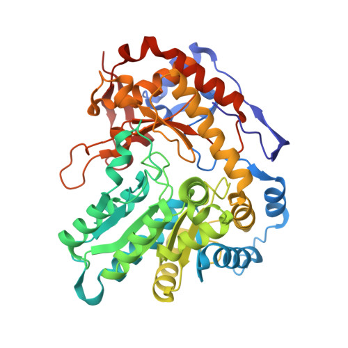
    8V6P, 8V9P, 9BLV, 9BNJ

  • PubMed Abstract: 

    Tryptophan indole lyase (TIL; [E.C. 4.1.99.1]) is a bacterial pyridoxal-5'-phosphate (PLP)-dependent enzyme that catalyzes reversible β-elimination of indole from L-tryptophan. The mechanism of elimination of indole from L-tryptophan starts with the formation of an external aldimine of the substrate and PLP, followed by deprotonation of the α-CH of the substrate, forming a resonance-stabilized quinonoid intermediate. Proton transfer to C3 of the indole ring and carbon-carbon bond cleavage of the quinonoid intermediate provide indole and aminoacrylate bound to PLP, which then releases indole, followed by iminopyruvate. We have now determined the X-ray crystal structures of TIL complexes with (3 S )-dioxindolyl-l-alanine, an inhibitor, and with substrates L-tryptophan, 7-aza-L-tryptophan, and S -ethyl-l-cysteine (SEC) in the presence of benzimidazole (BZI), an isostere of the product indole. These structures show a mixture of gem -diamine, external aldimine, quinonoid, and aminoacrylate intermediates, in both open and closed active site conformations. In the closed conformations of L-tryptophan, (3 S )-dioxindolyl-l-alanine, and 7-aza-L-tryptophan complexes, hydrogen bonds form between Asp-133 with N1 of the ligand heterocyclic ring and NE2 of His-458 in the small domain of TIL. This hydrogen bond also forms in the BZI complex with the aminoacrylate intermediates formed from both L-tryptophan and SEC. The closed quinonoid complex of 7-aza-L-tryptophan shows that the azaindole ring in the closed conformation is bent out of plane of the Cβ-C3 bond by about 40°, putting it in a geometry that leads toward the transition-state geometry. Thus, both conformational dynamics and substrate activation play critical roles in the reaction mechanism of the TIL.


  • Organizational Affiliation
    • Department of Chemistry, University of Georgia, Athens, Georgia 30602, United States.

Macromolecule Content 

  • Total Structure Weight: 212.33 kDa 
  • Atom Count: 16,188 
  • Modeled Residue Count: 1,864 
  • Deposited Residue Count: 1,868 
  • Unique protein chains: 1

Macromolecules

Find similar proteins by:|  3D Structure
Entity ID: 1
MoleculeChains  Sequence LengthOrganismDetailsImage
Tryptophanase
A, B, C, D
467Proteus vulgarisMutation(s): 0 
Gene Names: tnaA
EC: 4.1.99.1
UniProt
Find proteins for P28796 (Proteus vulgaris)
Explore P28796 
Go to UniProtKB:  P28796
Entity Groups
Sequence Clusters30% Identity50% Identity70% Identity90% Identity95% Identity100% Identity
UniProt GroupP28796
Sequence Annotations
Expand
Reference Sequence

Small Molecules

Ligands 3 Unique
IDChains Name / Formula / InChI Key2D Diagram3D Interactions
YQK
(Subject of Investigation/LOI)

Query on YQK


Download:Ideal Coordinates CCD File
G [auth A],
I [auth B],
K [auth C],
M [auth D]
(2~{E})-2-[(~{Z})-[2-methyl-3-oxidanyl-5-[[oxidanyl-bis(oxidanylidene)-$l^{6}-phosphanyl]oxymethyl]-1~{H}-pyridin-4-ylidene]methyl]imino-3-[(3~{S})-3-oxidanyl-2-oxidanylidene-1~{H}-indol-3-yl]propanoic acid
C19 H20 N3 O9 P
MNDDBLQWOZGYHE-LDSSDFKRSA-N
DMS

Query on DMS


Download:Ideal Coordinates CCD File
H [auth B]DIMETHYL SULFOXIDE
C2 H6 O S
IAZDPXIOMUYVGZ-UHFFFAOYSA-N
K

Query on K


Download:Ideal Coordinates CCD File
E [auth A],
F [auth A],
J [auth C],
L [auth D]
POTASSIUM ION
K
NPYPAHLBTDXSSS-UHFFFAOYSA-N

Experimental Data & Validation

Experimental Data

  • Method: X-RAY DIFFRACTION
  • Resolution: 1.85 Å
  • R-Value Free:  0.247 (Depositor), 0.247 (DCC) 
  • R-Value Work:  0.199 (Depositor), 0.199 (DCC) 
  • R-Value Observed: 0.201 (Depositor) 
Space Group: P 1 21 1
Unit Cell:
Length ( Å )Angle ( ˚ )
a = 67.342α = 90
b = 112.717β = 97.91
c = 119.518γ = 90
Software Package:
Software NamePurpose
PHENIXrefinement
Aimlessdata scaling
autoPROCdata processing
STARANISOdata scaling
XDSdata reduction
PHASERphasing

Structure Validation

View Full Validation Report


Ligand Structure Quality Assessment 


Entry History 

& Funding Information

Deposition Data


Funding OrganizationLocationGrant Number
National Institutes of Health/National Institute of General Medical Sciences (NIH/NIGMS)United StatesR01GM137008

Revision History  (Full details and data files)

  • Version 1.0: 2023-12-20
    Type: Initial release
  • Version 1.1: 2025-02-12
    Changes: Database references, Structure summary